Download
1 / 68
680 likes | 919 Vues
Hematology 425 Leukocyte Neoplasms. Russ Morrison November 29, 2006. Leukocyte Neoplasms. Neoplasm is defined as an abnormal tissue that grows by cellular proliferation more rapidly than normal and continues to grow after the stimuli that initiated the new growth ceases

E N D
Hematology 425 Leukocyte Neoplasms Russ Morrison November 29, 2006
Leukocyte Neoplasms • Neoplasm is defined as an abnormal tissue that grows by cellular proliferation more rapidly than normal and continues to grow after the stimuli that initiated the new growth ceases • A neoplasm may be benign as with a tumor or malignant in cancer(s) • A leukocyte neoplasm is abnormal cellular proliferation in the WBC series
Leukocyte Neoplasms • Leukemias are a group of diseases manifested by malignant, unregulated proliferation of cells normally found in the bone marrow • Leukemia may involve any of the blood-forming cells or their precursors • Leukemia may be found in metastatic lesions throughout the body, in the brain, liver, spleen and lymph nodes
Leukocyte Neoplasms • Though leukemias progress differently, the unregulated proliferating cells have four things in common • They usually replace normal marrow • They eventually interfere with normal marrow function • They may invade other organs • The eventually cause death if they go untreated
Leukocyte Neoplasms • Leukemias are relatively new diseases since they were first described in 1845 • In 1900, it was reported that acute and chronic leukemias involve different types of WBCs • In chronic leukemias, mature cells are predominant while in acute leukemias, immature cells (blasts) hold center stage
Leukocyte Neoplasms • Since the 1900s, leukemias have been studied with the microscope, cytochemical stains, electron microscopy, immunologic nuclear and surface markers and cytogenetic techniques • Standardized subclassification of leukemia subtypes leads to better treatments through pharmaceutical research • Today, we are at a threshold of development of targeted therapies, better treatments and cures for both the acute and chronic leukemias
Clinical Classification of Leukemia • Leukemias are divided into acute and chronic diseases based on their presenting signs and symptoms as well as the cell type involved • Acute leukemias are characterized by abrupt onset of clinical signs (infection, hemorrhage and pallor) and symptoms (fatigue, weakness, bone and joint pain) • Death occurs within months in acute leukemia if treatment is not begun
Clinical Classification of Leukemia • WHO classification of acute leukemias requires a minimum of 20% blast forms (myeloid or lymphoid) in either the PB or BM • PB WBC counts may be increased, decreased or normal, though typically they are increased • Normocytic anemia and neutropenia are classic states, while thrombocytopenia is usually present
Clinical Classification of Leukemia • Chronic leukemias have an insidious onset of signs (pallor, splenomegaly and/or hepatomegaly, weight loss) and symptoms (weakness, fatigue, depression) • They usually occur in adults and death occurs years after diagnosis • WBC counts are variable, though usually higher than seen in acute leukemia and cell forms are more mature • Platelet counts are normal or increased • Acute vs Chronic is compared in Table 32-1
Classification by Cell Type • A PB smear can initiate a diagnosis of acute or chronic leukemia • Additional information is gathered to subcategorize the lymphocytes in CLL and some of the CMLs using cytochemistry, flow cytometry, PCR and cytogenetics • Genetic testing to determine specific gene abnormalities will become standard within the next few years
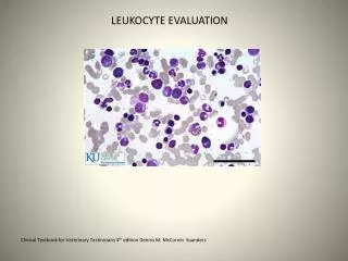
Lymphoblast 2-3 times the size of a normal lymphocyte Have scant blue cytoplasm Uniform coarse chromatin Inconspicuous nucleoli Myeloblast 3-5 times the size of a normal lymphocyte Moderate grey cytoplasm Uniform fine chromatin 2 or more prominent nucleoli Auer rods Morphologic identification of Lymphoblasts vs myeloblasts
Morphologic Subtypes of ALL • Three morphologic subtypes of “blasts” have been described according to the FAB classification system • Little, if any, prognostic information is gained from this classification • Subtypes are termed L1, L2 and L3 based on the following descriptions
ALL – L1 Lymphoblasts • Small • Scant blue cytoplasm • Round nuclei • Indistinct nucleoli • The most common type of ALL
ALL – L2 Lymphoblasts • 2-3 times the size of a normal lymphocyte • Moderate cytoplasm • Irregular nuclear membrane • Prominent nucleoli • May resemble AML
ALL – L3 Lymphoblasts • Large • Midnight blue cytoplasm • Cytoplasmic vacuoles are present • 3-5 nucleoli
Other Classifications of Acute Leukemias - Cytochemical • Cytochemical staining classification systems were developed by FAB • The current panel of stains includes myeloperoxidase, Sudan black B, chloroacetate esterase, and nonspecific esterase • Table 32-2 shows the FAB classification of acute leukemias by cytochemical reactions
Other Classifications of Acute Leukemias - immunologic • Monoclonal antibodies and flow cytometry added to morphology and cytochemical stains brings the accuracy of identification of leukemic cell lines to 95% • The technique is more commonly used for lymphoid rather than myelogenous cell lines • A panel of cell line markers is used because no single surface marker is specific • Table 32-3 shows the immunologic classification of these cells
Other Classifications of Acute Leukemias - Cytogenetic • Chromosomal alterations aree associated with leukemic cells • Cytogenetic studies can be used to detect clonal abnormalities • Clonal abnormalities can be of number (ploidy) or of structure (translocations, deletions and rearrangements)
Other Classifications of Acute Leukemias - Molecular • Molecular Diagnostics/PCR are able to identify genetic mutations in both acute and chronic leukemias • As these methods develop, the diagnosis and classification of acute and chronic leukemias will be performed by molecular methods
Acute Leukemias - ALL • Acute lymphoblastic leukemia (ALL) is a disease of childhood • Most cases occur between the ages of 2 and 10 years of age • All is rare in adults, but a second cluster of incidence occurs among elderly patients • Treatment of childhood ALL now yields a 90% rate of complete remission with a 60% cure rate
Acute Leukemias - ALL • Adults with ALL do not fare as well having a 68-91% remission rate and a 25-41% cure rate • Approximately half of patients with ALL have increased WBC counts and they may or may not have lymphoblasts in the PB • Neutropenia, thrombocytopenia and anemia are usually present • This pancytopenia causes fatigue, fever and bleeding and there is often lymph node enlargement
Acute Leukemias - ALL • Hepatomegaly and splenomegaly are often present as well as bone pain due to infiltration of leukemic cells into the periosteum • Eventually, the meninges are invaded by malignant cells and lymphoblasts are found in the CSF • Lymphoblast infiltration into the testes and ovaries is common, especially in relapse
ALL - Prognosis • Prognosis is dependent on the age of the patient at the time of diatnosis • It is also related to the lymphoblast load and immunophenotype • Chromosomal translocations (ex Philadelphia Chromosome 5(9;22)) are proving to be a strong predictor of adverse treatment outcomes for both children and adults
ALL - Prognosis • Patients 2-10 years of age have the best prognosis • Children less than 1 year of age do poorly • Children between 1 and 2 have an intermediate success rate, as do teenagers and young adults • Lymphoblast count > 20-30 x109/L, hepatosplenomegaly and lymphadenopathy all decrease the effectiveness of treatment and the patients are considered “high risk”
ALL - Prognosis • Other variables showing decreased outcomes include race (native American), karyotypic abnormalities and sex (male) • Morphology – • The three major FAB classifications FAB-L1, FAB-L2, and FAB-L3 were discussed in Chapter 32
ALL - Immunophenotyping • Morphology is the first tool in distinguishing ALL from AML, but immunophenotyping is the best indicator of a cell’s origin • Immunologic classification of ALL should be performed in all cases due to the prognostic implications
ALL - Treatment • Success with treatment is divided by age of presentation • Intrathecal methotrexate is used with children • Methotrexate is a folate antagonist and kids on this drug will be megaloblastoid • Adults are treated with daunorubicin, vincristine and prednisone (all with side effects) • Stem cell or bone marrow transplantation is the last line of treatment for unresponsive or relapsed ALL
Acute Myeloid Leukemia - AML • AML is the most common leukemia in children less than 1 year of age • AML is rare in older children and adolescents • A second age focus of AML occurs among adults 40 years of age • AML is rapidly fatal if not treated • 8 types of AML (M0 thru M7) have been described
AML – Clinical Presentation • Clinical presentation is nonspecific reflecting the decreased production of normal BM elements • WBC count is usually between 5000 and 30,000/mm3, but may be as high as 200,000 or as low as 1000 • Myeloblasts are present in the PB of 90% of patients • Anemia, thrombocytopenia and neutropenia lead to symptoms of pallor, fatigue, bruising, bleeding and fever with infections
AML – Clinical Presentation • DIC and other bleeding abnormalities occur, especially with M3-promyelocytic and monocytic-M5 forms of AML • Infiltration of malignant cells into the gums and mucous membranes, as well as the skin are is seen in M4-myelomonocytic and M5-monocytic types • Bone and joint pain is the first symptom in 25% of patients • Splenomegaly is seen in half of AML patients, but lymph node enlargement is rare
AML – Clinical Presentation • AML patients do not usually exhibit symptoms when the CNS is infiltrated with blasts • Elevated uric acid, potassium and phosphate levels along with decreased calcium (termed tumor-lysis syndrome), renal failure, tetany and lethal heart arrythmias may develop
AML – Subtypes • Micrographs of the AML subtypes are exhibited in the text and atlas • The FAB cooperative group has defined 8 subtypes (M0-M8) of AML based on bone marrow morphology and cytochemical reactions • In the FAB classification, 2 types of myeloblasts have been described • Type 1 have typical blast morphology and no azurophilic granules • Type 2 may contain a small number of primary granules
AML – Subtype M0 • Acute myeloblastic leukemia, minimally differentiated • Blasts do not display morphologic myeloid characteristics • Fewer than 3% of blasts exhibit myeloperoxidase or Sudan black B positivity • Auer rods are not sen • May express antigen TdT
AML – Subtype M1 • Acute myeloblastic leukemia, without maturation • Displays minimal myeloid differentiation • Myeloblasts constitute more than 30% of the BM nucleated cells • M:E ratio is greater than 1 • 90% of the erythroid cells are myeloblasts • 3% of the myeloblasts show positivity when stained with myeloperoxidse of Sudan black B • Easily confused with ALL (L2 type) • Accounts for 20% of cases of AML
AML – Subtype M2 • Acute myeloblastic leukemia, with maturation • 30% of nucleated cells must be blasts in the BM film and myeloid cells outnumber NRBCs • Blasts constitute fewer than 90% of nonerythroid cells and there is maturation beyond the promelycyte in more than 10% of nonerythroid cells • Majority of blasts stin positively when stained with myeloperoxidase or Sudan black B • Subtype accounts for 30% of cases of AML
AML • Induction therapy in M1 and M2 leukemia is usually effective, resulting in sufficient return of normal marrow responses for an autologous bone marrow transplant • Drugs frequently used are daunorubicin and cytosine arabinoside • Cardiac monitoring is necessary due to side effects • Cytosine arabinoside is active against the nucleus, resulting in a megaloblastoid presentation of the CBC
AML – Subtype M3 • Acute promyelocytic leukemia • May be one of two types, hypergranular or microgranular • Stain strongly positive with myeloperoxidase • M3 patients must be recognized because bleeding problems (DIC) occur when these patients are treated and the granules are released • Make up approximately 15% of all AML cases • Treatment with all-trans retinoic acid (ATRA) a form of Vitamin A causes the cells to undergo maturation (cells have a defective vitamin A receptor)
AML – Subtype M4 • Acute myelomonocytic leukemia • Has malignant cells with both granulocytic and monocytic features • BM more than 30% of nucleated cells are blasts • When the M:E ratio is greater than 1, more than 20% of nonerythroid cells are monocytic • A small percent has moderate eosinophilia (AML-M4Eo), have CSF involvement and a better remission rate and response to chemotherapy
AML – Subtype M5 • Acute monocytic leukemia • Accounts for 12% of AML • Diagnosis based solely on the BM morphology • More than 30% of cells are blasts • More than 80% of cells are monocytic, less than 20% granulocytic • Two subtypes recognized, M5a and M5b • Skin involvemen and gum infiltration may be seen in both types
AML – Subtype M6 • Acute erythroleukemia • Rare, 3% of AML cases • The only AML with hyperplasia of erythroid precursors
AML – Subtype M7 • Acute megakaryocytic leukemia • The rarest type of AML at <1% of cases • In adults, many cases are preceded by a myeloproliferative disorder with pancytopenia or myelofibrosis • In children it is more frequent among patients under three years of age with Down syndrome • Response to therapy is poor
Other Acute Myeloid-like Leukemias • Acute undifferentiated leukemia and hybrid leukemias are often treated as AML, but are not necessarily related • Undifferentiated acute leukemia is a rare disorder in which the lineage of the blasts can not be determined • Hybrid leukemias are leukemias with both lymphoid and myeloid characteristics
Chronic leukemias – lymphocytic disorders • There are two major types of chronic lymphoid leukemias • Chronic lymphocytic leukemia (CLL) • Hairy cell leukemia There are also a few other forms of chronic lymphoid leukemias that are rare and not discussed in this series
Chronic Lymphocytic Leukemia • CLL is the most common type of leukemia • Usually occurs in older patients • Most commonly found in patients older than 40 (though youngest case was 22) • Affects twice as many men as women • No specific chromosomal abnormality has been associated with CLL