1 / 59

How to read X’Ray chest

Dr. S. Parthasarathy MD., DA., DNB, MD ( Acu ), Dip. Diab . DCA, Dip. Software statistics Ph d (physiology) Mahatma Gandhi medical college and research institute , puducherry – India . How to read X’Ray chest. Preliminary check up . Is it the right patient ?

merry
Télécharger la présentation

How to read X’Ray chest

An Image/Link below is provided (as is) to download presentation Download Policy: Content on the Website is provided to you AS IS for your information and personal use and may not be sold / licensed / shared on other websites without getting consent from its author. Content is provided to you AS IS for your information and personal use only. Download presentation by click this link. While downloading, if for some reason you are not able to download a presentation, the publisher may have deleted the file from their server. During download, if you can't get a presentation, the file might be deleted by the publisher.

E N D

Presentation Transcript


  1. Dr. S. Parthasarathy MD., DA., DNB, MD (Acu), Dip. Diab. DCA, Dip. Software statistics Ph d (physiology) Mahatma Gandhi medical college and research institute , puducherry – India How to readX’Raychest

  2. Preliminary check up • Is it the right patient ? • Is it the right position ? • Overall patient ? Gopal or ramu

  3. XRay interprétation

  4. Rotation • Medial end of the clavicle – equidistantfromvertebra – • closerlungappearswhiter Clavicle Clavicle vertebra

  5. Left rotation

  6. Exposure • Check at the lower part of the cardiacshadow • Spinesjust visible – normal • Cleardemarcation of spines – overpenetration • Spines not visible – underpenetration

  7. Over and under penetration

  8. Or demarcation at T4

  9. Apex missed

  10. Rt CP angle ??

  11. Inspiration • 6th rib anterior • 9th rib posterior • If no – • Heart larger • Basal shadows • Trachea to right

  12. Things to see • Soft tissue • Bones • Mediastinum • Angles • Heart and vessels • Lungs • Go methodical

  13. Soft tissue Emphysema

  14. bones - Scapula

  15. Bones – ribs

  16. Scoliosis

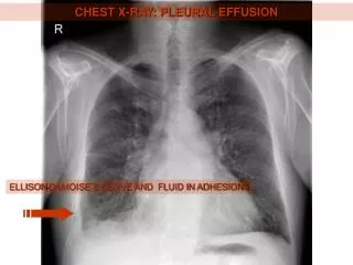
  17. Costophrenic angles • Rt diaphragm lower • 1 inch • Highest point – rt – middle of the lung field • Lt – slightly lateral • Smooth contour

  18. CP angles which is low and why?

  19. Mediastinum- fuzziness Aorta Pulm. art LA LV

  20. CT ratio 50 %

  21. Prominent aortic knuckle

  22. Pulmonary hypertension

  23. Left atrial and LV enlargement

  24. SVC and rt atrium enlarged

  25. Where is the trachea ?

  26. MEDIASTINUM

  27. Lung parenchyma • Rotation • Exposure • Soft tissue • Bones • Mediastinum • Trachea • Angles

  28. Pneumonia

  29. Pneumothorax - markings ??

  30. Collapse • Upper zone- from 2nd costal cartilage to axilla • Middle zone- between 2nd and 4th costal cartilage. • Lower zone- below 4th costal cartilage

  31. Active PT ?

  32. Silicosis

  33. Perforation

  34. Emphysema

  35. Pneumonectomy

  36. Some life lines can be assessed • Central line • Tracheostomy • Nasogastric tube • ET tube

  37. Tracheal tube position

  38. Central lines

  39. Nasogastric tube

  40. Pacing wires

  41. Reading an X-ray

  42. How to read

  43. Rt sided chest pain and cough

  44. Cough fever

  45. Two X rays

  46. Students turn

  47. Post op laparotomy

  48. Silhoute sign ??

  49. What is it??

  50. Smoker for years dry cough wheezing

More Related