Download
1 / 17
170 likes | 309 Vues
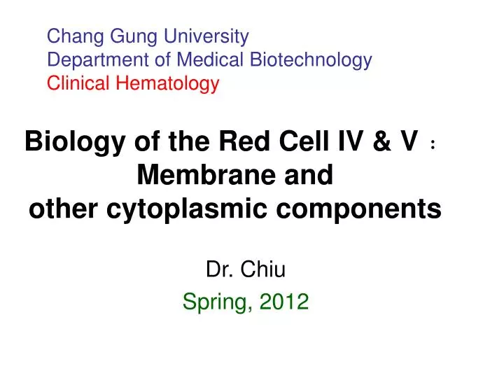
Chang Gung University Department of Medical Biotechnology Clinical Hematology. Biology of the Red Cell IV & V﹕ Membrane and other cytoplasmic components. Dr. Chiu Spring, 2012. Lecture Outline. I. The red cell membrane structure

E N D
Chang Gung UniversityDepartment of Medical Biotechnology Clinical Hematology Biology of the Red Cell IV & V﹕ Membrane and other cytoplasmic components Dr. Chiu Spring, 2012
Lecture Outline I. The red cell membrane structure A. Membrane structural components﹕ Lipids, proteins and carbohydrates B. The bilayer coupling hypothesis C. Asymmetry of membrane phospholipids II. Functions of the red cell membrane A. Maintenance of cell volume 1. Na+ and K+ content 2. Osmotic fragility B. Ca+2 Homeostasis C. Anion exchange
Lecture Outline V. Required reading Text (Essential Haematology): pp 18-20. VI. References 1. Rifkind r, et al: Fundamentals of Hematology, pp55-56,71-73, 1986 2. Lux, S. and S. Shohet. The erythrocyte membrane skeleton. Hospital Practice pp 77-83, Oct. 1984 3. Lubin B, Kuypers F, Chiu D. Red cell membrane lipid dynamices in The Red Cell. Alan R. Liss, Inc. 1989 pp507-524 4. Beutler, E. Red cell metabolism: A manual of biochemical methods. Crune and Stratton, 1984 5. Chiu, DTY, Liu TZ. Free radical and oxidative damage in human blood cells. J Biomed. Sci 4: 256-259, 1997.
Lecture Outline I. The red cell membrane structure A. Membrane structural components﹕ Lipids, proteins and carbohydrates B. The bilayer coupling hypothesis C. Asymmetry of membrane phospholipids II. Functions of the red cell membrane A. Maintenance of cell volume 1. Na+ and K+ content 2. Osmotic fragility B. Ca+2 Homeostasis C. Anion exchange
Biological Membranes as Bilayer Couples. A Molecular Mechanism ofDrug-Erythrocyte Interactions Sheetz MP, Singer SJ. Proc Natl Acad Sci U S A. 1974 Nov;71(11):4457-61.
ABSTRACT We propose that membranes whose proteins and polar lipids are distributed asymmetrically in the two halves of the membrane bilayer can act as bilayer couples, i.e., the two halves can respond differently to a perturbation. This hypothesis is applied to the interactions of amphipathic drugs with human erythrocytes. It is proposed that anionic drugs intercalate mainly into the lipid in the exterior half of the bilayer, expand that layer relative to the cytoplasmic half, and thereby induce the cell to crenate, while permeable cationic drugs do the opposite and cause the cell to form cup-shapes. This differential distribution of the drugs is attributed to interactions with the phosphatidylserine that is concentrated in the cytoplasmic half of the membrane. Impermeable amphipathic drugs intercalate only into the exterior half of the bilayer, and therefore are crenators of the intact cell. Several predictions of this hypothesis have been confirmed experimentally with erythrocytes and erythrocyte ghosts. The bilayer couple hypothesis may contribute to the explanation of many membrane-mediated phenomena in cell biology.
Membrane bilayer coupling with changing RBC morphology
A schematic representation of the red cell membrane structure Sphinomyeline (SM) Phosphatidylcholine (PC) Phosphatidylethanoline (PE) Phosphatidylserine (PS)
C. Asymmetry of membrane phospholipids
Lecture Outline I. The red cell membrane structure A. Membrane structural components﹕ Lipids, proteins and carbohydrates B. The bilayer coupling hypothesis C. Asymmetry of membrane phospholipids II. Functions of the red cell membrane A. Maintenance of cell volume 1. Na+ and K+ content 2. Osmotic fragility B. Ca+2 Homeostasis C. Anion exchange
Lecture Outline III. Red cell deformability ( A brief review ) A. Factors affecting red cell deformability 1. Cytoplasmic viscosity 2. intracellular rubbish 3. membrane rigidity 4. surface to volume ratio B. Techniques to measure red cell deformability 1. filtration 2. micropipette 3. Ektacytometry (Vidometry) IV. Other cytoplasmic components and red cell metabolism A. Glucose metobolism﹕ glycolysis and the hexomonophosphate shunt B. Protective mechanisms against oxidative damage
Lecture Outline III. Red cell deformability ( A brief review ) A. Factors affecting red cell deformability 1. Cytoplasmic viscosity 2. intracellular rubbish 3. membrane rigidity 4. surface to volume ratio B. Techniques to measure red cell deformability 1. filtration 2. micropipette 3. Ektacytometry (Vidometry) IV. Other cytoplasmic components and red cell metabolism A. Glucose metobolism﹕ glycolysis and the hexomonophosphate shunt B. Protective mechanisms against oxidative damage
Glucose metabolism • Luebering-Rapoport shunt Fig 2.10 • Embden-Meyerhof glycolytic pathway • Hexose monophosphate shunt pathway
Oxidative stress and the protective mechanism Oxy Hb Met Hb In red cells, there is a Met Hb reductase which can use NADH to regenerate Hb
Protective systems in cells against peroxidative reactions NADPH H2O ROH GSSG Hexose Monophosphate Shunt(G6PD) Glutathione Peroxidase Glutathione Reductase Superoxide Dismutase Catalase O2 H2O2 ROOH GSH NADP G-6-P Met Hb Reductase to regenerate Hb using NADH Glutathione Synthetase Fe3+ Amino Acid OH Vit. E ( oxid. Vit. E can be recycled by Vit. C ) Damage Cellular components leading to accelerated cell removal