Download
1 / 11
110 likes | 320 Vues
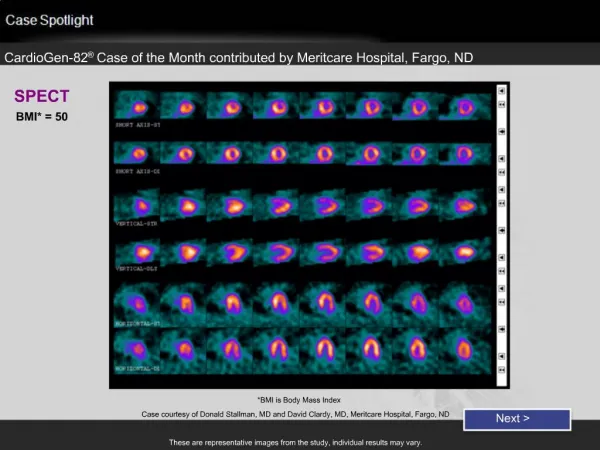
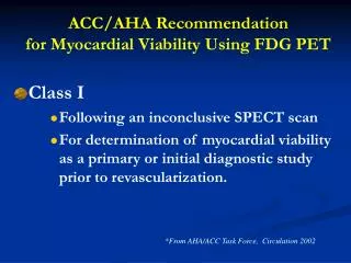
ACC/AHA Recommendation for Myocardial Viability Using FDG PET. Class I Following an inconclusive SPECT scan For determination of myocardial viability as a primary or initial diagnostic study prior to revascularization. *From AHA/ACC Task Force, Circulation 2002.

E N D
ACC/AHA Recommendation for Myocardial Viability Using FDG PET • Class I • Following an inconclusive SPECT scan • For determination of myocardial viability as a primary or initial diagnostic study prior to revascularization. *From AHA/ACC Task Force, Circulation 2002
Recommendations for detection of myocardial viability in heart failure Hunt SA, et al. Circulation 2005;112:e154–e235. Underwood SR, et al. Eur Heart J 2004;25:815–836.
Criteria for acute MI • Detection of elevated values ofcardiac biomarkers (preferably troponin) above the 99th centileof the upper reference limit (URL) together with evidence ofmyocardial ischaemia with at least one of the following: • Ischaemicsymptoms • ECG changes indicative of new ischaemia (new ST-Tchanges or new left bundle branch block (LBBB)) • Developmentof pathological Q waves in the ECG • Imaging evidence of newloss of viable myocardium or new regional wall motion abnormality. Thygesen, K. et al. Circulation 2007;116:2634-2653
ZXL, 67-year-old man with angina and no prior MI, 90% stenosis of LAD , 60% stenosis of LCx He ZX, et al. Circulation 2003
71-y-old man (patient 7) with exertional angina, 90% stenosis of LAD and 80% stenosis of LCx and RCA Dou KF, Yang MF, et al. J Nucl Med 2008 ; 49:1986–1991
Imaging of Ischemic Memory With BMIPP Dilsizian, V. et al. Circulation 2005;112:2169-2174.
BMIPP SPECT Imaging in a Patient With Suspected ACS (Kontos MC, et al. J Am Coll Cardiol 2010;56:290–9)
ROC Curves for Patients Imaged <12 h Vs. 12 to 30 h After Symptom Resolution (Kontos MC, et al. J Am Coll Cardiol 2010;56:290–9)
Nuclear imaging procedures in the United States in 2007 3% ↑ 5% ↑
Relative frequency of procedures by organ or System in Beijing in 2005 Bone Lung Kidney Cardiovascular Total procedure, 84734 Si HW, et al. NMC 2007