Download
1 / 32
350 likes | 434 Vues
Dive into the evaluation of a patient with acute chest pain and shortness of breath, emphasizing pulmonary embolism diagnosis and radiological chest anatomy. Understand chest imaging findings and differential diagnoses through interactive learning.

E N D
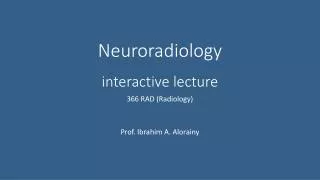
Interactive CHEST IMAGINGlecture 366 COURSE October 2016
Question ? • Patient presented to ER with acute chest pain and SOB • Past history of pelvic fracture with hospital admission for 5 weeks. • Answer the following question by selecting the SINGLE BEST ANSWER 1. ------------------- 2.-------------------- X 3.-------------------- 4.--------------------
Answer ? • Patient presented to ER with acute chest pain and SOB • Past history of pelvic fracture with hospital admission for 5 weeks. PULMONARY EMBOLISM Q-SINGLE BEST ANSWER 1. ------------------- 2.CTA showing PULMONARY EMBOLISM X 3.-------------------- 4.--------------------
trachea Anatomy Costophrenic angle Diaphragmatic cupola
Radiological Anatomy of the Chest L R Upper Lobe Upper Lobe ????? ????? Lower Lobe Major Fissure Lower Lobe Major Fissure LEFT LUNG RIGHT LUNG
Radiological Anatomy of the Chest L R Upper Lobe Upper Lobe Minor Fissure Middle Lobe Lower Lobe Major Fissure Lower Lobe Major Fissure LEFT LUNG RIGHT LUNG
LUNG WINDOW Consolidation
DIAGNOSIS • 37 YEARS OLD PATIENT WITH SOB AND FEVER.
MASS OR INFILTERATATION ? STEPS 1 2 3 ---?--- WINDOW
EXAM? ?
EXAM? CALCIFIC PLAQUE ? CT ANGIOGRAPHY CORONARY ARTERIES CATHETER ANGIOGRAPHYCORONARY ARTERIES
HEART ?
HYDRO-PNEUMOTHORAX AIR FLAT FLUID LEVEL FLAT FLUID LEVEL