Download
1 / 121
1.21k likes | 1.56k Vues
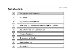
Table of Contents – pages iii. Unit 1: What is Biology? Unit 2: Ecology Unit 3: The Life of a Cell Unit 4: Genetics Unit 5: Change Through Time Unit 6: Viruses, Bacteria, Protists, and Fungi Unit 7: Plants Unit 8: Invertebrates Unit 9: Vertebrates Unit 10: The Human Body.

E N D
Table of Contents – pages iii Unit 1:What is Biology? Unit 2:Ecology Unit 3:The Life of a Cell Unit 4:Genetics Unit 5:Change Through Time Unit 6:Viruses, Bacteria, Protists, and Fungi Unit 7:Plants Unit 8:Invertebrates Unit 9:Vertebrates Unit 10:The Human Body
Table of Contents – pages vii-xiii Unit 1: What is Biology? Chapter 1:Biology: The Study of Life Unit 2: Ecology Chapter 2:Principles of Ecology Chapter 3:Communities and Biomes Chapter 4:Population Biology Chapter 5:Biological Diversity and Conservation Unit 3:The Life of a Cell Chapter 6:The Chemistry of Life Chapter 7:A View of the Cell Chapter 8:Cellular Transport and the Cell Cycle Chapter 9:Energy in a Cell
Unit 4: Genetics Chapter 10:Mendel and Meiosis Chapter 11:DNA and Genes Chapter 12:Patterns of Heredity and Human Genetics Chapter 13:Genetic Technology Unit 5: Change Through Time Chapter 14:The History of Life Chapter 15:The Theory of Evolution Chapter 16:Primate Evolution Chapter 17:Organizing Life’s Diversity Table of Contents – pages vii-xiii
Unit 6: Viruses, Bacteria, Protists, and Fungi Chapter 18:Viruses and Bacteria Chapter 19:Protists Chapter 20:Fungi Unit 7: Plants Chapter 21:What Is a Plant? Chapter 22:The Diversity of Plants Chapter 23:Plant Structure and Function Chapter 24:Reproduction in Plants Table of Contents – pages vii-xiii
Table of Contents – pages vii-xiii Unit 8: Invertebrates Chapter 25:What Is an Animal? Chapter 26:Sponges, Cnidarians, Flatworms, and Roundworms Chapter 27:Mollusks and Segmented Worms Chapter 28:Arthropods Chapter 29:Echinoderms and Invertebrate Chordates
Table of Contents – pages vii-xiii Unit 9: Vertebrates Chapter 30:Fishes and Amphibians Chapter 31:Reptiles and Birds Chapter 32:Mammals Chapter 33:Animal Behavior Unit 10: The Human Body Chapter 34:Protection, Support, and Locomotion Chapter 35:The Digestive and Endocrine Systems Chapter 36:The Nervous System Chapter 37:Respiration, Circulation, and Excretion Chapter 38:Reproduction and Development Chapter 39:Immunity from Disease
Unit Overview – pages 890-891 The Human Body Protection, Support, and Locomotion The Digestive and Endocrine System The Nervous System Respiration, Circulation, and Excretion Reproduction and Development Immunity from Disease
Chapter Contents – page xiii Chapter 34Protection, Support, and Locomotion 34.1:Skin: The Body’s Protection 34.1:Section Check 34.2:Bones: The Body’s Support 34.2:Section Check 34.3:Muscles for Locomotion 34.3:Section Check Chapter 34Summary Chapter 34 Assessment
Chapter 32 Chapter Intro-page 892 What You’ll Learn You will interpret the structure and functions of the integumentary system. You will identify the functions of the skeletal system. You will classify the different types of muscles in the body.
32.1 34.1 Section Objectives – page 893 Section Objectives: • Compare the structures and functions of the epidermis and dermis. • Identify the role of the skin in responding to external stimuli. • Outline the healing process that takes place when the skin is injured.
32.1 Section 34.1 Summary – pages 893-898 Structure and Functions of the Integumentary System • Skin, the main organ of the integumentary (inh TE gyuh MEN tuh ree) system, is composed of layers of the four types of body tissues: epithelial, connective, muscle, and nervous. • Epithelial tissue, found in the outer layer of the skin, functions to cover surfaces of the body.
32.1 Section 34.1 Summary – pages 893-898 Structure and Functions of the Integumentary System • Connective tissue, which consists of both tough and flexible protein fibers, serves as a sort of organic glue, holding your body together. • Muscletissues moves parts of the body. In the skin, muscle interacts with hairs on the skin to respond to stimuli, such as cold and fright.
32.1 Section 34.1 Summary – pages 893-898 Structure and Functions of the Integumentary System • Nervous tissue helps us detect external stimuli, such as pain or pressure. Epidermis Dermis
32.1 Section 34.1 Summary – pages 893-898 Structure and Functions of the Integumentary System • Skin is composed of two principal layers—the epidermis and dermis. Epidermis Dermis
32.1 Section 34.1 Summary – pages 893-898 Epidermis: The outer thinner layer of skin
32.1 Section 34.1 Summary – pages 893-898 Epidermis: The outer thinner layer of skin • The top layer of cells, although dead & flattened, serves an important function as they contain a protein called keratin (KER uh tun). • Keratin helps to waterproof and protect the living cell layers underneath from exposure to bacteria, heat, and chemicals.
32.1 Epidermis: The outer thinner layer of skin Section 34.1 Summary – pages 893-898 • The interior layer of the epidermis contains living cells that continually divide by mitosis to replace the dead cells. • Some of these cells contain melanin, a pigment that colors the skin and helps protect body cells from damage by solar radiation. Even though the # of melanin-producing cells is about the same in each person, the amt. of melanin produced per cell varies, resulting in different colors of skin. • Every four weeks, all cells of the epidermis are replaced by new cells.
32.1 Section 34.1 Summary – pages 893-898 Epidermis: The outer thinner layer of skin • The epidermis on the fingers and palms of your hands, and on the toes and soles of your feet, contain ridges and grooves that are formed before birth. • These epidermal ridges are important for gripping as they increase friction.
32.1 Dermis: The inner thicker layer of skin Section 34.1 Summary – pages 893-898 The thickness of this layer varies depending on the function of that body part.
32.1 Oil glands Section 34.1 Summary – pages 893-898 The Skin Hair The fat layer is below the dermis. It functions to store E, provide insulation, & acts a shock absorber. Sweat glands
32.1 Functions of the integumentary system Section 34.1 Summary – pages 893-898 • Primary function is PROTECTION • One function of skin is to help maintain homeostasis by regulating your internal body temperature. • Erector pili muscle & goose bumps – forma a layer of insulation to prevent heat loss. • When your body temperature rises, the many small blood vessels in the dermis dilate, blood flow increases, and body heat is lost by radiation.
32.1 Section 34.1 Summary – pages 893-898 Functions of the integumentary system • When you are cold, the blood vessels in the skin constrict and heat is conserved. • Glands in the dermis produce sweat in response to an increase in body temperature. • As sweat evaporates, water changes state from liquid to vapor and heat is lost.
32.1 Section 34.1 Summary – pages 893-898 Functions of the integumentary system 3 functions of the oil produced by the skin: • Keeps the hair moist • Keeps the skin soft & pliable • Inhibits the growth of some bacteria
32.1 Section 34.1 Summary – pages 893-898 Functions of the integumentary system • Skin also functions as a sense organ. • Nerve cells in the dermis receive stimuli from the external environment and relay information about pressure, pain, and temperature to the brain.
32.1 Section 34.1 Summary – pages 893-898 Functions of the integumentary system • Another function of the skin is to maintain a • chemical balance of certain substances, such • as Vitamin D. • When exposed to ultraviolet light, skin cells produce vitamin D, a nutrient that aids the absorption of calcium into the bloodstream.
32.1 Section 34.1 Summary – pages 893-898 Functions of the integumentary system • Cuts or other openings in the skin surface allow bacteria to enter the body, so they must be repaired quickly.
32.1 Section 34.1 Summary – pages 893-898 Skin Injury and Healing • When the epidermis sustains a mild injury, such as a scrape, the deepest layer of epidermal cells divide to help fill in the gap left by the abrasion. • If, however, the injury extends into the dermis, where blood vessels are found, bleeding usually occurs.
32.1 Section 34.1 Summary – pages 893-898
32.1 Section 34.1 Summary – pages 893-898 Skin Injury and Healing • Burns can result from exposure to the sun or contact with chemicals or hot objects. • Burns are rated according to their severity.
32.1 Section 34.1 Summary – pages 893-898 Skin Injury and Healing • First-degree burns, such as a mild sunburn, involve the death of epidermal cells and are characterized by redness and mild pain. • First-degree burns usually heal in about one week without leaving a scar.
32.1 Section 34.1 Summary – pages 893-898 Skin Injury and Healing • Second-degree burns involve damage to skin cells of both the epidermis and the dermis and can result in blistering and scarring. • The most severe burns are third-degree burns, which destroy both the epidermis and the dermis. • With this type of burn, skin function is lost, and skin grafts may be required to replace lost skin.
32.1 Section 34.1 Summary – pages 893-898 Skin Injury and Healing • As people get older, their skin changes. • It becomes drier as glands decrease their production of lubricating skin oils—a mixture of fats, cholesterol, proteins, and inorganic salts.
32.1 Section 34.1 Summary – pages 893-898 Skin Injury and Healing • Wrinkles may appear as the elasticity of the skin decreases.
32.1 Section 1 Check Question 1 What part of the skin responds to external stimuli such as heat and pressure? A. epithelial tissue B. nervous tissue C. connective tissue D. muscle tissue
32.1 Section 1 Check The answer is B, nervous tissue. Nerve endings
32.1 Section 1 Check Question 2 Why is your skin considered an organ? Answer Skin is composed of cells and tissues. A group of tissues that work together to perform a specialized function are called an organ.
32.1 Section 1 Check Question 3 The thicker portion of skin that is composed of blood vessels, nerves, nerve endings, hair follicles, sweat glands, and oil glands is called the _______. A. dermis B. subcutaneous layer C. epidermis D. fat tissue
32.1 Section 1 Check The answer is A, dermis.
32.2 Section 2 Objectives – page 899 Section Objectives • Compare the different types of movable joints. • Describe how bone is formed. • Identify the structure and functions of the skeletal system.
32.2 Section 34.2 Summary– pages 899-904 Skeletal System Structure • The adult human skeleton contains about 206 bones. • Its two main parts are shown.
32.2 Section 34.2 Summary– pages 899-904 Skeletal System Structure
32.2 Section 34.2 Summary– pages 899-904 Joints: Where bones meet • In vertebrates, joints are found where two or more bones meet. • Most joints facilitate the movement of bones in relation to one another. • The joints of the skull, on the other hand, are fixed joints, as the bones of the skull don’t move.
32.2 Section 34.2 Summary– pages 899-904
32.2 Section 34.2 Summary– pages 899-904 Joints: Where bones meet • Joints are often held together by ligaments. • A ligament is a tough band of connective tissue that attaches one bone to another. • In movable joints, the ends of bones are covered by cartilage. • This layer of cartilage allows for smooth movement between the bones.
32.2 Section 34.2 Summary– pages 899-904 Joints: Where bones meet • In addition, joints such as those of the shoulder and knee have fluid-filled sacs called bursae located on the outside of the joints. • The bursae act to decrease friction and keep bones and tendons from rubbing against each other.
32.2 Section 34.2 Summary– pages 899-904 Joints: Where bones meet • Tendons, which are thick bands of connective tissue, attach muscles to bones. • Forcible twisting of a joint, called a sprain, can result in injury to the bursae, ligaments, or tendons. • A sprain most often occurs at joints with large ranges of motion such as the wrist, ankle, and knee.
32.2 Section 34.2 Summary– pages 899-904 Joints: Where bones meet • One common joint disease is arthritis, an inflammation of the joints. • One kind of arthritis results in bone spurs, or outgrowths of bone, inside the joints. • Such arthritis is especially painful, and often limits a person’s ability to move his or her joints.
32.2 Section 34.2 Summary– pages 899-904 Compact and spongy bone • Bones are composed of two different types of bone tissue: compact bone and spongy bone. Spongy bone Marrow cavity Humerus Compact bone Periosteum Artery Vein
32.2 Section 34.2 Summary– pages 899-904 Compact and spongy bone • Surrounding every bone is a layer of hard bone, or compact bone. Spongy bone Marrow cavity Humerus Compact bone Periosteum Artery Vein