Download
1 / 26
260 likes | 407 Vues
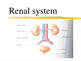
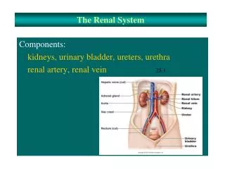
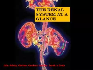
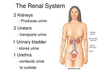
The Renal System. 2 Kidneys - Produces urine. 2 Ureters - transports urine. 1 Urinary bladder - stores urine. 1 Urethra - conducts urine to outside. 3 Layers Surrounding the Kidney. Renal Capsule - inner layer lines renal sinus. Adipose Capsule - fat padding around kidney.

E N D
The Renal System 2 Kidneys - Produces urine 2 Ureters - transports urine 1 Urinary bladder - stores urine 1 Urethra - conducts urine to outside
3 Layers Surrounding the Kidney Renal Capsule - inner layer lines renal sinus Adipose Capsule - fat padding around kidney Renal Fascia - outermost layer
Outer renal cortex Inner renal medulla Central renal pyramid Renal papilla Minor calyx Major calyx Renal pelvis Ureter
Calyx – ‘cup’ Pelvis = basin Calyx - singular Calicies - plural
There are 6-18 renal pyramids in a kidney: - The renal papillae project into renal sinus. Renal lobe - Contains renal pyramid, overlying cortex, adjacent renal column. Collection of Urine Minor calyx Major calyx Renal Pelvis (continuous with ureter)
Renal artery • Segmental artery • Interlobar artery • Arcuate artery • Interlobular artery • Afferent arteriole Veins carry same names
The Nephron has 2 parts: 1. Renal Corpuscle Glomerulus Bowman’s Space Bowman’s Capsule 2. Renal Tubule Proximal convoluted tubule (PCT) Loop of Henle (nephron loop) Distal convoluted tubule (DCT)
~85% Cortical Nephrons Short loop of Henle. Efferent arteriole becomes peritubular capillaries. 15% Juxtamedullary Closer to medulla. Loop of Henle deeper. Efferent arteriole becomes vasa recta.
Juxtaglomerular Apparatus Macula Densa – specialized portion of DCT. Sits in between afferent and efferent arteriole – for communication regarding renal function. Juxtaglomerular (JG) Cells – specialized cells, mostly surrounding afferent arteriole. For changing diameter of afferent arteriole.
Glomerulus – Fenestrated Capillary bed • Blood arrives at afferent arteriole. Filtrate “filtered” at Glomerulus. • Blood departs via efferent arteriole. • Blood goes into a) Peritubular or b) Vasa recta capillaries • Blood exits via renal venule, vien.
Proximal Convoluted Tubule • Actively reabsorbs • Nutrients (glucose, amino acids) • Ions • Small Plasma proteins • Lined with simple cuboidal epi • With microvilli (brush border) • Large surface area
Loop of Henle Descending limb Ascending limb • Thin segment is simple squamous epi. • Thick segment is simple cuboidal epi (no nicrovilli) • For Water balance Distal Convoluted Tubule Actively secretes ions and other materials Lined with simple cuboidal epi.
Collecting Duct Lined with simple cuboidal epi. Changes to simple columnar epithelium deeper in the medulla.
Bladder is lined with . . . Transitional epithelium