Download
1 / 1
10 likes | 139 Vues
T he phytohormone resveratrol and the pineal hormone melatonin i n the rat mammary carcinogenesis.

E N D
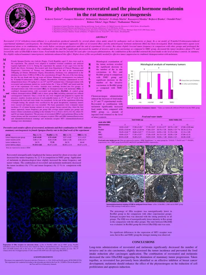
The phytohormone resveratrol and the pineal hormone melatonin in the rat mammary carcinogenesis Kisková Terézia*1, Garajová Miroslava1, Böhmdorfer Michaela2, Svoboda Martin3, Kassayová Monika1, Bojková Bianka1, Orendáš Peter1, Bobrov Nikita4, Jäger Walter2, Thalhammer Theresia3 1. Department of Animal Physiology, Institute of Biology and Ecology, Faculty of Science, P.J. Šafárik University, Moyzesova 11, 040 01 Košice, Slovak Republic. 2. Department of Clinical Pharmacy and Diagnostics, University of Vienna, Althanstrasse 14, A-1090 Vienna, Austria. 3. Department of Pathophysiology, Center for Pathophysiology, Infectiology & Immunology, Medical University of Vienna, Währinger Gürtel 18-20, 1090 Vienna, Austria. 4. Department of Forensic Medicine, Faculty of Medicine, P.J. Šafárik University, Trieda SNP 1, 040 11 Košice, Slovak Republic. ABSTRACT Resveratrol (3,5,4’-trihydroxy-trans-stilbene) is a phytoalexin produced naturally by several plants when under attack by pathogens such as bacteria or fungi. In a rat model of N-methyl-N-nitrosourea-induced experimental mammary carcinogenesis, the chemopreventive and tumor-suppressive effect of resveratrol (Res) and the antiestrogenic hormone melatonin (Mel) has been investigated. Both chemopreventive agents were administered alone or in combination, two weeks before carcinogen application until the end of experiment (18 weeks). Res alone slightly lowered tumor frequency in comparison with other groups and prolonged the latency period for about seven days. The combination of Res and Mel significantly decreased the number of invasive and in situ carcinomas as compared to NMU group, decreased the tumor incidence about 17% and lowered the ratio of ERα/ERβ in tumor tissue. Food intake declined in 2nd and 7th week after carcinogen administration; the combination of Res and Mel returned the food intake to the level of intact controls. In summary, Res slightly reduced mammary tumorigenesis, melatonin enhanced this effect. Higher doses of Res are probably needed for effective tumor suppression in combined chemoprevention with melatonin. M A T E R I A L S and M E T H O D S Female Sprague-Dawley rats (AnLab, Prague, Czech Republic) aged 31 days were used in the experiment. The animals were adapted to standard vivarium conditions and artificial regimen light:dark 12:12 h. During the experiment the animals (4 per cage) were fed the Ssniff diet (Soest, Germany). Resveratrol (gift from Department of Clinical Pharmacy and Diagnostics, University of Vienna) was administered in the dietat the concentration 100mg/kg ad libitum. Melatonin (Sigma, Deisenhofen, Germany) was administered in the drinking water from 15:00h to 8:00h at the concentration 20 mg/l. The rest of the time during the day the rats drank only the tap water ad libitum. Mammary carcinogenesis was induced by N-methyl-N-nitrosourea (NMU) (Sigma, Deisenhofen, Germany) administered in two intraperitoneal doses (50 mg/kg b.w.) on 43rd and 50th postnatal days. Chemoprevention with resveratrol and melatonin started two weeks before the 1st dose of NMU and lasted until the end of the experiment (18 weeks). Animals were randomly divided into five groups: a) chemoprevention only with resveratrol (Res), b) chemoprevention with melatonin (Mel), c) combined chemoprevention with resveratrol and melatonin (ResMel), d) control group without chemoprevention (NMU) and e) intact group (Int)including untreated rats without carcinogenesis induction.Once a week the rats were weighed and palpated to register the presence, number, location and size of each palpable tumor; in 2nd, 7th and 12th week of the experiment the food and water intake was monitored. At the end of the experiment following overnight fasting, the animals were sacrificed by the quick decapitation, mammary tumors were excised and tumor size was recorded. The basic parameters were evaluated: tumor incidence (% of tumor bearing animals in every group), latency period (day, when the first tumor appeared), tumor frequency per group (the average tumor number per group) and tumor volume (calculated according to the formula V= π x (S1)2 x S2/12; S1< S2, where S1 and S2 are tumor diameters). The presence of resveratrol (HPLC) and melatonin (Elisa) in the serum plasma and the assessment of estrogen receptors ERα and ERß (immunofluorescence and immunohistochemical staining) and melatonin receptor MT1 (immunohistochemical staining) were determined. Histological examination of the tumor sections revealed the significant decrease in the number of the in situ carcinomas in Res and ResMel group in comparison with NMU group and significant decrease in the number of the invasive carcinomas in ResMel group as compared with NMU group. R E S U LTS Chemocarcinogen administration induced the decrease of food intake in 2nd and 7th experimental weeks. Resveratrol in combination with melatonin significantly increased food intake when compared with NMU group, i.e. amount of ingested food returned to the level of intact controls. Histological analysis of mammary tumors. * Values are statistically different (P<0,05) from the NMU group. Food and water intake Preventive and curative effects of resveratrol, melatonin and their combination in NMU- induced mammary carcinogenesis in female Sprague-Dawley rats in the final week of the experiment Food and water intake per rat.Data are expressed as means ± SEM. Significance * vs. intact group (by * P<0,05; ** P<0,01 and *** P<0,001, respectively)● vs. NMU group (by ● P<0,05; ●● P<0,01 and ●●● P<0,001, respectively). b Data are expressed as means ± SEM. a Resveratrol nonsignificantly lengthened the latency period for about seven days and decreased the tumor frequency by 22 % in comparison to NMU group. Application of melatonin in pharmacological dose slightly increased the tumor frequency and tumor volume. The combination of resveratrol and melatonin moderately decreased the tumor incidence (by 17%) and tumor frequency (by 13 %) in comparison with NMU. a b c d Immunohistochemical staining of ERβ in malignant breast tissue. a) Res, b) ResMel, c) Mel and d) NMU group. Brown ERβ staining of milk ducts is shown. c The percentage of ERα receptors was nonsignificantly lower in ResMel group in the comparison with other experimental groups. Estrogen receptor beta was detected with the strong positivity in all groups. The ERβ was of nonsignificantly lower intensity in Res group in the comparison with the other groups. The ratio between ERα/ERβ was evaluated. In ResMel group the lowest ERα/ERβ ratio was seen. No significant differences in the expression of MT1 receptor were seen, but in Res and NMU group the stronger staining was observed. d CONCLUSIONS Long-term administration of resveratrol and melatonin significantly decreased the number of invasive and in situ carcinomas, slightly decreased the tumor incidence and prevented the food intake reduction after carcinogen application. The combination of resveratrol and melatonin decreased the ratio ERα/ERβ suggesting the diminution of mammary tumor progression. Taken together, as resveratrol has previously been identified as an effective inhibitor of breast cancer development, melatonin should enhance the effect of the phytoestrogen on the reduction of cell proliferation and apoptosis induction. Expression of ERα receptor in cancerous tissue. a) Res b) ResMel c)Mel and d) NMU group. Paraffin embedded sections from breast tissue were stained for ERα by immunofluorescence. The pictures in 1st column show all DAPI stained cells in the layer, in 2nd column the cells expressing ERα receptors are seen. The 3rd column shows the overlapped picture of cells stained with DAPI and cells expressing ERα (FITC). ACKNOWLEDGEMENTS The project was supported by Liga proti rakovine; Chemosvit, a.s. Svit; SAIA and OeAD agency (ICM-2009-02358). The experiment was conducted according to the principles provided in the Law No. 23/2009 of Slovak Republic for the Care and Use of Laboratory Animals.