Download
1 / 1
40 likes | 426 Vues
Hemodynamic Analysis of Arteriovenous Fistula Configurations. Briana Conners , Chemical Engineering, University of Cincinnati & Ken echuwku Okoye , Biomedical Engineering, University of Cincinnati Graduate Mentor : Ehsan Rajabi Jaghargh , Mechanical Engineering, University of Cincinnati

E N D
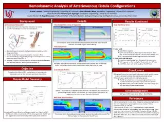
Hemodynamic Analysis of Arteriovenous Fistula Configurations Briana Conners, Chemical Engineering, University of Cincinnati & Ken echuwkuOkoye, Biomedical Engineering, University of Cincinnati Graduate Mentor: EhsanRajabiJaghargh, Mechanical Engineering, University of Cincinnati Faculty Mentor: Dr. RupakBanerjee, Professor, School of Dynamic Systems ,College of Engineering and Applied Science, University of Cincinnati • Results • Results Continued Axial Wall Shear Stress Flow Streamlines: Areas of Stagnation • Background • Inner bend: • Axial WSS is negative • By the end of the bend, WSS return to the direction of the flow on the inner bend in the 90° case but not in the 60° case • 60° case experiences higher negative levels of axial WSS • Outer bend and Side bend: • Experience positive axial WSS throughout the entire curve • WSS is greater on the first half of the bend for the 60° case • Axial WSS values vary less throughout the 90° bend • Arteriovenous Fistula (AVF) • Connection of the end of a vein to the side of an artery. • Used for hemodialysis in terminal renal patients. • Blood pathway circumvents the high-resistance capillaries in the distal extremities • Vessels must dilate to accommodate the increase in flow • Vessel remodeling is triggered by changes in hemodynamics, namely the shear stress on the wall [2] • However, 23-46% of all fistula do not achieve an adequate diameter and resulting flow as a result of venous stenosis [1] Cells lining the blood vessel wall become disoriented from flow not in the axial direction. This likely triggers wall thickening. [3] Axial Velocity Profiles • Objective Section 1: axial velocity is negative for the inner half. The negative flow velocity is of greater magnitude in the 60° case than the 90° case and even continues to be negative on the inner curve at the end of the bend. • Conclusions • Stagnant flow on the inner bend is observed in both models in areas in which stenosis formation has been documented before. • The 90° case is preferred for the following reasons: • The 60° case had more pronounced stagnant flow on the inner bend which may result in more prominent stenosis • High WSS on the outer wall of the 60 ° case may damage wall cells and lead to further complications Velocity Contours • Fistula Model Geometry To explore the effects of AVF configuration on hemodynamic parameters such as flow field and wall shear stress (WSS). Along the outer bend of the fistula, velocity magnitude is greater than on the inner bend. The highest velocity fluid is noted on the outer wall of the 60° case. • Acknowledgement NSF Type 1 STEP Grant, Grant ID No.: DUE-0756921 Wall Shear Stress Contours 1.Krishnamoorthy, M. K. et al., 2012, “Anatomic configuration affects the flow rate and diameter f porcine arteriovenous fistulae.” Kidney International, 81, pp. 745-750. 2.Ene-Iordache, B. et al., 2011, “Disturbed flow in radial-cephalic arteriovenous fistulae for haemodialysis: low and oscillating shear stress locates the sites of stenosis.” Nephrol Dial Transplant, 27, pp. 358-368. 3.“What is Arterio-Venous Access.” Cardiac Vascular and Thoracic Surgery Associates. Web. Nov. 2012. <http://www.cvtsa.com/ListofConditions/A-444-176.html.>. • References Using blood flow and blood vessel data averaged from 6 porcine AVF, the fistula models were created with the above geometry at a 90° and 60 ° angle to be solved numerically under steady state condition. The friction of the flowing blood creates a shear stress on the walls of the vessels. The WSS are higher on the side wall of the 60° case.