Hemodynamic Disorders
Hemodynamic Disorders. Vascular dynamics I- Blood flow – Normal fluid homeostasis 1- Edema 2- Hyperemia 3- Hemorrhage II- Maintainence of blood as a liquid 1- Hemostasis 2- Thrombosis III- Embolism IV- Infarction V- Shock.

Hemodynamic Disorders
E N D
Presentation Transcript
Vascular dynamics I- Blood flow – Normal fluid homeostasis 1- Edema 2- Hyperemia 3- Hemorrhage II- Maintainence of blood as a liquid 1- Hemostasis 2- Thrombosis III- Embolism IV- Infarction V- Shock
1- Normal fluid homeostasis - Intact circulation - Maintenance of vessel wall integrity - Physiologic ranges - Intravascular pressure - Osmolarity 2- Altered vascular homeostasis results in : - Change in net movement of water across the vascular wall
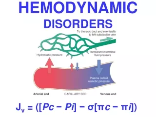
Edema = Excess accumulation of fluid in the interstitial tissue spaces or body cavities . - Under normal circumstances only a small amount of fluid leaks from vessels to form interstitial fluid which is removed by lymphatic vessels. - Causes : more fluid leaves capillaries than enters : 1- If the hydrostatic pressure in vessels is increased ( e.g., interference with venous drainage , congestive heart failure) 2- Decreased plasma oncotic pressure ( hypoproteinemia – albumin) a- Nephrotic synd. - loss of protein in kidney b- Decreased albumin production in liver during cirrhosis 3- Vascular permeability is altered ( allergic responses liberate histamine , acute inflammation , burn injury
4-Increased sodium retention - Primary – assoc. with renal disorders - Secondary – occurs in congestive heart failure - Decreased cardiac output …….decreased renal blood flow ……activation of renin-angiotensin system …….aldosterone activated ……retention of sodium & water. 5- Blockage of lymphatics ; results in lymphedema Causes of Edema : 1- Increased permeability 2- Increased hydrostatic pressure 3- Decreased oncotic pressure 4- Lymphatic obstruction
Two important types of edema due to cardiac failure : I- Pulmonary edema 1- Accumulation of fluid in the lung alveoli 2- Caused by increased hydrostatic pressure in the pulmonary vascular bed resulting from left – side heart failure II- Peripheral ( subcutaneous ) edema 1- Accumulation of fluid in subcutaneous tissues 2- Caused by increased hydrostatic pressure in the systemic venous system resulting from right – side heart failure
Types of edema : 1- Anasarca : generalized edema 2- Hydrothorax : accumulation of fluid in pleural cavity 3- Hydropericardium : Abnormal accumulation of fluid in the pericardial cavity which may result in cardiac tamponade 4- Hydroperitoneum ( ascites) : Abnormal accumulation of fluid in the peritoneal cavity 5- Transudate : Non-inflammatory edema fluid that results form altered intravascular hydrostatic pressure or osmotic pressure ; due to low protein content & specific gravity < 1.012 6- Exudate :Edema fluid from increased vascular permeability as a result of inflammation: High protein content & specific gravity > 1.020 , Contains large number of inflammatory leukocytes which often consume glucose & thus results in glucose content being greatly reduced.
Hyperemia • Localized increase in the volume of blood in capillaries & small vessels • - Active hyperemia : results from localized arteriolar dilation ( blushing , inflammation
Hyperemia - passive congestion ( passive hyperemia) results from obstructed venous return or increased back pressure from CHF 1 - Active passive congestion – shock , acute inflammation , sudden right heart failure 2- Chronic passive congestion : - Lung – left heart failure or mitral stenosis cause ; congestion , distension of alveolar capillaries ……..rupture …….heart failure cells ( hemosiderin laden macrophages) - Liver & lower extremities – right heart failure cause with nutmeg liver ( combination of dilated congested central veins , & brown yellow fatty liver cells)
HYPEREMIA Active Process CONGESTION Passive Process Acute or Chronic
Hemorrhage : Escape of blood from the vasculature into surrounding tissues , hollow organ or body cavity or to the outside - Caused by rupture of blood vessels - Massive exsanguination usually caused by trauma to a major artery or vein but may also be from bursting of a vessel weakened by disease - Bleeding into tissues or body cavities results in several types of hemorrhage
Types of hemorrhage : - Hematoma : accumulation of blood within soft tissues usually due to trauma of vessels but occasionally follows spontaneous rupture of diseased vessels. - Petechia & Ecchymosis : 1-2mm & -10 mm , respectively tissue hemorrhages of the skin or oral mucosa due to abnormal small vessel fragility , abnormal blood clotting or abrupt increase in pressure within small venules & capillaries - Hemopericardium : Collection of blood in the pericardial cavity due to rupture of the heart or the aorta ; may result in cardiac tamponade.
- Hemothorax : Collection of blood in the pleural cavities due to trauma or rupture of the aorta - hemoperitoneum : Collection of blood in the peritoneum due to rupture of an aortic aneurysm or trauma to liver , spleen , or aorta - Hemoarthrosis : Collection of blood in a joint space due to trauma or bleeding disorder ( e.g. haemophilia)
Hemostasis - Normal hemostasis results from well regulated processes that maintain blood in a fluid , clot – free state in normal vessels while inducing the rapid formation of a localized hemostatic plug at the site of vascular injury. - Dependent on the vascular wall , platelets & the coagulation cascade ( as is pathological thrombosis) - Has a normal general sequence of events
Thrombosis : Process of thrombus formation due to activation of the normal blood coagulation system - An intravascular coagulation of blood often causing significant interruption of blood flow - Predisposed by venous stasis , CHF , polycythemia , sickle cell disease , visceral malignancies , oral contraceptives esp. when combined with cigarette smoking A thrombus is a structured solid mass composed of blood constitutes ( platelets , insoluble fibrin , embedded RBCs) that forms within the cardiovascular system N.B. : not a coagulum = unstructured & forms when blood clots outside the circulatory system.
Morphological characteristics : -Arterial thrombi - Formed in areas of active blood flow - Mature have dark layers of platelets interspersed with lighter layers of fibrin = lines of Zahn = Laminated layers of platelets & fibrin/RBCs - Eventually liquefy & disappear or organized with fibrous tissue formation
Venous thrombi ( phlebothrombosis) - Form in areas of less active blood flow , most often veins of the lower extremities & periprostatic or other pelvic veins - Predisposed to venous stasis -Dark red with highter concentration of RBCs than arterial thrombi so lines of Zahn not present or not prominent - Often associated with concurrent venous inflammatory changes.
Hemostasis / thrombosis Coagulation cascade : - Ultimate aim is to generate a solid plug of cross lined protein that seals a defect in a blood vessel ; protein deposited is fibrin generated from it is circulating precursor protein fibrinogen - To achieve this aim many different protein interact in a cascade : - Each coagulation factor has a number 1-XIII - Nearly all of these are functionally proteases ; factors V & VIII are not ; act as co-factors
Hemostasis / Thrombosis Compartments of the coagulation cascade - Common pathway : - Results in cross – linked fibrin - Thrombin is the key protease - Has feedback to activate co-factors , other proteases & thus amplifies the cascade - Extrinsic pathway : - Coagulation initiated by tissue factor ( generated by damaged tissue) interacting with factor VII - Intrinsic pathway : - Coagulation initiated by contact with surface agents ( e.g. collagen , kallikrien ) acting through factor XII ( Hageman factor) - Currently through to have minor role for in vivo coagulation - Activation of factory XI & coagulation stimulation is seen mainly after severe injury ( e.g. trauma)
Details of the intrinsic pathway - Coagulation initiated by tissue factor generated on cells surface adjacent to vessels & exposed following injury to the vessel wall ; tissue factor + VIIa …. Activate IX & also X - Ixa + VIIIa & Ca+ act on platelet phospholipid ( PPS) …..X ……XA ; VIII = parts C ( coag. Pathway & vWF [ co-factor activated by thrombin ]) - Xa ….. Complex on pps with Va & Ca+ …..prothrombin …..thrombin - Thrombin cleaves fibrinogen into fibrin & fibrinopeptides A, B - Thrombin activates XIII……crosslinkage of fibrin …thrombus - Thrombin activates XI , VIII , V - Thrombin acts on endothelial cells & promotes vasoconstrictive factors & plasminogen activator
- Products of the coagulation cascade usually restricted to site of vessel wall damage - Plasma inhibitors limit the cascade - Antithrombin III is most potent especially via action of heparin - Protein C : - Vitamin K dependent - Activated by thrombin + thrombomodulin + protein S ….. Destroy Va & VIIIa - Allows fibrinolysis - Fibrinolysis : Due to formation plasminogen ….. Proteas plasmin via plasminogen activators , tPA & uPA [ no longer inhibited by plasminogen activator inhibitor 1 but protein C prevents this ] …..degrades fibrin …..fibrinopeptides ( fibrin degradation products with anticoagulant activity )
- Thrombosis events ( thrombogenesis) – results from interction of platelets , damaged endothelial cells & the coagulation cascade - Aggregation of platelets held together with a meshwork of fibrin occurs constantly to plug small defects in blood vessel walls ; once vessel wall repaired the small platelet / fibrin thrombus is normally removed via fibrinolysis ; multienzyme process that destroys fibrin filament meshwork allowing dissolution of the thrombus - Excessive thrombosis is prevented by several physiological mechanisms but in pathological thrombosis the thrombus formation proceeds beyond the capacity of the endogenous fibrinolysins to eradicate the thrombus …….thrombus enlarges by deposition of fresh layers ( laminated) of platelets & fibrin until lumen of vessels may be reduced.
Thrombosis : The normal endothelial cells act to prevent activation of the coagulation cascade generating factors that bring about fibrin lysis : - Intact endothelium prevents platelets from contacting collagen & von Willebrand factor ( cause platelet aggregation & degranulation) - Prostacyclin & nitric oxide prevent adhesion & aggregation of platelets to endothelium - Thrombomodulin on the endothelium surface binds to local fibrin formed ……thrombomodulin / thrombin complex initiates anticoagulant effects of vitamin K dependent factor protein C & it is cofactor protein S ( active protein C destroys factors V & VIII)
Normal endothelial cells - Produces heparin – like molecules which inhibit elements of the cascade - Synthesizes plasminogen activators which produce plasmin …..lyses fibrin & inactivates part of the cascade - Heparin potentiates antithrombin III a potent inhibitor of coagulation
Three main factors predisposes to thrombus formation 1- Endothelial dysfunction : direct injury ( trauma & inflammation ; atheroma) 2- Changes in the flow pattern of blood : stasis allows platelets to come into contact with endothelium & prevents dilution of activated coagulation products . 3- Changes in the potential blood coagulability : - Increase in the conc. Of fibrinogen in acute phase responses - Congenital lack of protein C , protein S , antithrombin III - Antiphospholipid antibodies - Leiden mutation – factor
- Thrombosis in different parts of the circulation have different causative factors & different macroscopic appearances - Fast moving blood in arteries & heart chambers have high platelet / fibrin content so are very firm , pale , prominent laminations - Slow moving venous blood have a high proportion of entrapped RBCs relative to platelet / fibrin so are red , soft , gelatinous with poor laminations - Occlusive thrombi ; small & medium sized vessels completely occluded - Mural thrombi ; in the heart or aorta without complete occlusion i.e. vegetations – thrombi on heart valves.
- 4 main outcomes of occlusive thrombi 1- Propagation : May enlarge along the vessel or undergo lysis by the fibrinolytic system 2- Organization : ingrowth of granulation tissue from vessel wall 3- Recanalization : Gradual replacement by granulation tissue & new vascular channels develop bridging the site of occlusion & re- establising flow 4- Thromboembolism : Fragments break of thrombus & carried by the circulation to impact other vessels
Embolism - Occlusion of a vessel by a mass of material ( i.e. embolus ) that is transported in the blood stream - Most common type due to fragments of circulating thrombus b( thromboemboli) - Fragments break off site of formation to enter blood circulation where it travels until it meets a blood vessel with a lumen too small to permit further passage: - If in systemic veins ……heart …..pulmonary thromboembolism - If in heart …..aorta ……systemic arterial …..arteries of brain , kidneys , spleen , gut & lower limbs - Common carotid arteries …..cerebral arteries - Abdominal aorta …….renal arteries & arteries of L.L.
Pulmonary thromboembolism - Most common preventable cause of sudden death in a hospital patient - Most commonly caused by thrombosis of deep leg vein ( calf , popliteal , femoral , iliac veins ) ……pulmonary artery ( saddle embolus = straddles bifurcation ) …….hemorrhage pulmonary infarct
- Other sources of pulmonary thromboemboli : very rarely peri – prostatic venous plexus in males ; small pelvic veins in women - Clinical predisposers : Immboility & bed rest ; postoperative period , pregnancy & post partum , oral contraceptives with high estrogen , nephrotic syndrome , severe burns , trauma , cardiac failure , disseminated malignancy - Two main consequences : - Increase in pulmonary arterial pressure ( strains right side of heart) - Ischemia of the lungs