Download
1 / 29
420 likes | 1.29k Vues
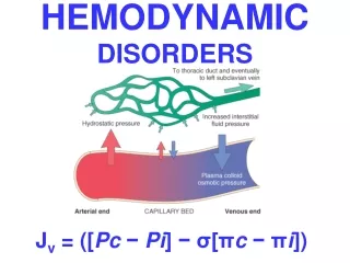
Hemodynamic Disorders, Thrombosis, Shock. Matthew Velkey matt.velkey@duke.edu 454A Davison, Duke South, Green Zone. Fluid Homeostasis. Total extracellular tissue fluid volume = hydrostatic pressure – [ colloid osmotic pressure + lymphatic drainage ].

E N D
Hemodynamic Disorders, Thrombosis, Shock Matthew Velkey matt.velkey@duke.edu 454A Davison, Duke South, Green Zone
Fluid Homeostasis Total extracellular tissue fluid volume = hydrostatic pressure – [colloid osmotic pressure + lymphatic drainage] Disruption in fluid homeostasis = edema
Localized Edema • Inflammatory • Combination of increased blood flow due to arteriolar vasodilation AND increased leakiness of capillary endothelium • Mechanical • Blockage of lymph vessels • Filariasis (nematode infection) • Neoplasia • Chemoterapy/radiotherapy damage to lymphatics
Systemic Edema • Increased hydrostatic pressure • Gravity • Congestive Heart Failure • Venous Obstruction • DVT, Vena cava obstruction • Cirrhosis –backs up blood in hepatic portal system • Constrictive Pericarditis –similar to CHF, heart can’t pump • Reduced Osmotic Pressure • Liver failure (not making enough albumin) • Nephrotic syndrome (losing too much albumin) • Sodium (and water) retention • Acute renal failure
Pathways to systemic edema Reduced Oncotic Pressure • Malnutrition • Liver failure/dysfunction • Can’t make enough albumin • Also causes hepatic portal congestion • Nephrotic syndrome • Glomerular capillaries too leaky and albumin is lost in urine
Pathways to systemic edema Congestive Heart Failure • RV failure: blood backs up in vena cava • LV failure: blood backs up first in lungs, then vena cava • Low cardiac output stimulates Renin-Angiotensin-Aldosterone pathway and sets up vicious cycle in kidneys • AngII raises systemic BP • Aldosterone increases Na+ retention • ADH increases water retention
Edema in Congestive Heart Failure Right-sided Heart Failure: Right atrial pressure Systemic venous pressure Congestion where? Increases Increases Systemic - Centrilobular Liver Congestion (venous) - Lower Extremity Edema
Centrilobular Hepatic Congestion Blood backs up in liver and impedes flow of oxygenated blood into deepest zones (around central veins), thus causing “central” necrosis and giving the liver a mottled, “nutmeg” appearance.
Edema in Congestive Heart Failure Left-sidedHeart Failure: Left atrial pressure: Pulmonary venouspressure: Pulmonary edema/congestion: Key feature of left heart failure, can lead to pulmonary edema Right atrial pressure: Increases Increases No initial change
Chronic Pulmonary Congestion Blood extravasates from capillaries and is taken up by resident macrophages that become engorged with hemosiderin at which point they are commonly referred to as “heart failure cells.”
Hemorrhage In skin, mucous membranes, or serosal surfaces: Petechiae - tiny (1-2 mm) Purpura- medium-sized (> 3 mm & < 1 cm) Ecchymoses - bruises (> 1 cm) Hematoma = collection of blood in an organ or tissue: Hemothorax: in the thorax Hemoperitoneum: in the peritoneum Hemopericardium: in the pericardium Hemarthrosis: in joint
Hemostasis Sequence of events following vascular injury that results in the formation of a clot (stasis) Key regulators are endothelial cells and platelets
Platelets (thrombocytes) • Life Span: about 10 days • Shape, size, and origin: Small, biconvex disks, 2-3 µm in diameter. Non-nucleated cell fragments derived from cytoplasm of a very large cell, the megakaryocyte, in bone marrow. Platelets have a life span of about 10 days. • LM appearance in smears: Small basophilic fragments, often appearing in clusters. • TEM appearance: The platelet is bounded by a plasma membrane, and has a bundle of microtubules around the margin of the disk (which maintains the disk shape). There are three types of granules, containing fibrinogen, plasminogen, thromboplastin and other factors for clotting. There are also membrane tubules and glycogen. • Function: Platelets initiate blood clots.
Transmission electron micrographs of a platelet seen in cross section (above) and in a section in the plane of the disk (below) granule membrane tubule
Cutaway diagram of a platelet • Peripheral microtubule bundle (maintains shape) • Actin and myosin (clot contraction) • Organelles facilitate clotting: • Mitochondria for ATP production • Granules contain clotting factors • Dense tubular system sequesters Ca++ for signaling (similar to SR in skeletal muscle) • Open canalicular system facilitates signaling and secretion (Ca++)
thromboplastin thrombin Fibrin polymerization Prothrombin Fibrinogen Thrombin Fibrin Ca++ Platelets and blood clot formation When a blood vessel wall is damaged, factors from the damaged endothelial cells and the ECM induce the clotting cascade. Platelets aggregate and release proteins for clot formation and resolution: 1. Vasoconstriction –via release of endothelin (from endothelium) 2. Further platelet aggregation –mediated via thromboxane A2and ADP 3. Fibrin polymerization –initiated by thromboplastin and free Ca++ 4. Clot contraction –via actin, myosin, and ATP released into the matrix of the clot 5. Clot resolution –platelet plasminogen activator(pPA, converts plasminogen into active fibrinolyticplasmin) 6. Tissue repair –platelet derived growth factor(PDGF, stimulates smooth muscle and fibroblast proliferation)
Endothelial modulation of clotting Antithrombotic properties • Antiplatelet effects: • Endothelial prostacyclin (PGI2) and Nitric Oxide inhibit platelet aggregation • ADPase: degrades ADP thus inhibiting platelet aggregation • Anticoagulant effects: • Heparin-like co-factors mediate antithrombin III inactivation of thrombin • Thrombomodulin binds and converts thrombin to an anticoagulant enzyme that activates protein (which then inactivates downstream clotting factors) • Fibrinolytic effects: • tissue plasminogen activator (tPA) activates plasmin which promotes lysis of clots
Endothelial modulation of clotting Prothrombotic properties • von Willebrand Factor: • Cofactor made by endothelial cells and bound to underlying collagen; when exposed allows platelets to bind to collagen and start to aggregate • Tissue factor: • Activates clotting cascade • Induced by proinflammatory cytokines such as IL-1 and TNF • Plasminogen activator inhibitors (PAIs): • Preventscleavage of plasminogen into active plasmin, thus inhibiting fibrinolysis
Perturbations in hemostasis result in thrombosis • Endothelial injury: • Direct injury • Depletion of anticoagulants (e.g. PGI2 by Cox-2 inhibitors) • Upregulation of procoagulants (e.g. inflammation) Virchow’s triad • Abnormal blood flow: • Aneurismal dilation of vessels create local stasis • Hyperviscosity (too many erythrocytes in blood) • Sickle cell anemia • Turbulence at branchpoints • Hypercoagulability: • Protease-resistant clotting factors (e.g. factor V Leiden): resistant to cleavage and therefore more active • Hormonal: estrogen increases production of clotting factors and reduces anticoagulant factors • Heparin-induced thrompocytopenia: administration of full-length heparin causes antibodies to develop that inactivate its antithrombotic activities • Antiphospholipid antibody syndrome: often seen in autoimmune disease (e.g. lupus), Abs activate platelets and inhibit PGI2 synthesis, thus promoting hypercoagulable state.
Fate of a thrombus • Dissolution: fibrinolytic activity completely clears thrombus • Organization and recanalization or incorporation: thrombi in vessels induce inflammation and fibrosis (organization); these can recanalize (shown below) or they can become incorporated into the vessel wall • Propagation: thrombus stimulates further platelet aggregation and growth that may eventually occlude vessel lumen • Embolization: thrombi may break off and plug a distant site
Venous vs. arterial thromobosis Venous thrombosis • Superficial (varicosities): cause local edema, pain, and perhaps ulceration; rarely embolize • Deep (i.e. “DVT”): rarely cause local pain due to collaterals, but often embolize with significant consequences Arterial thrombosis • Atherosclerosis: rupture of plaques induces clotting and occlusion of vessels • Mural thrombosis: post-infarction or post-infection damage to lining of heart induces formation of clots that can break off and plug a distant site.
Embolism Embolus = detached mass that is carried to a site distant from its origin, for example: Fat: bone marrow or soft tissue trauma releases adipocytes into blood that can plug distant sites Air: rapid depressurization causes gas to bubble out of solution; these bubbles block blood vessels causing infarction in muscles, brain, and other organs Amniotic fluid: trauma during childbirth may allow amniotic fluid (and its non-fluid contents such as dead skin cells, mucus, etc.) to enter maternal circulation and cause remote blockages.
Thromboembolism Embolism causing blockage is derived from a thrombus Pulmonary thromboembolism: thrombus (usually from a DVT) breaks off and goes to right ventricle. From there it is pumped out to the lungs and blocks pulmonary arteries. The problem at first is not the ischemia per se, but instead that this blood is not oxygenated and does not return to the heart (thus eventually causing systemic ischemia). Systemic thromboembolism: thrombus originates in left ventricular wall or wall of aorta breaks off and causes infarction at a distant site (brain, kidney, spleen).
Infarction Area of ischemic necrosis caused by occlusion of arterial supply or venous drainage. The nature and extent of damage is influenced by: Nature of blood supply: tissues with dual or collateral blood supply (e.g. lungs, liver, and limbs) are less affected compared to end organs (muscles, brain, kidney, spleen) Rate of development: slowly progressing occlusion tolerated because of development of collateral routes Tissue vulnerability:neurons can withstand only 3-4 minutes of hypoxia, myocytes ~30 minutes, fibroblasts can survive many hours in low oxygen.
Shock Systemic hypoperfusion caused by reduced cardiac output or reduced blood volume, resulting in 1) hypotension, 2) impaired tissue perfusion, and 3) cellular hypoxia. General categories are: Cardiogenic: reduced cardiac output Hypovolemic: reduced blood volume Hemorrhagic: blood loss Neurogenic: vasodilation following nerve cord injury Anaphylactic: systemic vasodilation and respiratory insufficiency Septic: over-reactive inflammatory response to infection
Septic shock Cytokine storm of TNF, IL-1, and IL-6 in response to bacterial antigens (usu. lipopolysaccharides from gram-negative bacilli) resulting in: • Systemic vasodilation • Reduced cardiac contractility • Widespread endothelial injury and activation • Systemic activation of clotting cascade (Disseminated Intravascular Coagulation or “DIC”)
General stages of shock • Nonprogressive stage: reflex compensatory mechanisms (tachycardia, peripheral vasoconstriction, renal fluid retention) compensate for hypoperfusion. • Causes common symptoms associated with shock: weak, rapid pulse; shallow, rapid breathing; and cool, clammy skin (the exception is septic shock that may present with flushing due to widespread inflammatory response). • Progressive stage: metabolic lactic acidosis blunts vasomotor response and blood starts to pool in peripheral tissues (increasing hypercoagulative risk), vital organs perfused less and begin to fail. • Clinical symptoms associated with this phase are reduced urine output, acidosis, and electrolyte imbalances • Irreversible stage: widespread tissue necrosis induces systemic inflammatory response (vasodilation, etc.), reduced cardiac function, and acute renal failure.