Download

1 / 39
490 likes | 1.55k Vues
WELLONE PRIMARY MEDICAL AND DENTAL CARE For Medical Provider Staff. Vaginal pH and Wet Mount Testing. Click here to move on. WARNING. Conducting this test exposes the operator to potentially infectious material. Standard precautions, including glove use are required for this procedure.

E N D
WELLONE PRIMARY MEDICAL AND DENTAL CARE For Medical Provider Staff Vaginal pH and Wet Mount Testing Click here to move on
WARNING Conducting this test exposes the operator to potentially infectious material. Standard precautions, including glove use are required for this procedure. Click here to go back Click here to move on
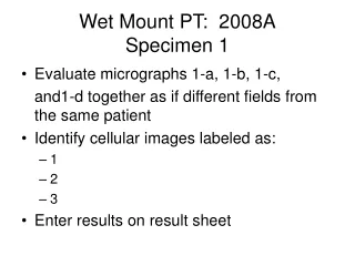
Patients with symptoms of bacterial vaginosis generally complain of an increased vaginal discharge with a foul, fishy odor that becomes more pronounced with menstruation or after unprotected intercourse. The patient may also experience some attendant irritation. Examination discloses a normal vulvar area and vagina as well as a copious, homogeneous, white-to-gray discharge, which is easily wiped from the vaginal wall. Ph Testing and Wet Mount • Vaginal pH testing and wet mount are indicated based on patient complaints and symptoms; rarely is “asymptomatic” pH or wet mount testing indicated. • The absence of trichomonads or pseudohyphae does not rule out infections because several studies have demonstrated the presence of these pathogens by culture or PCR after a negative microscopic examination. Click her to go back Click here to move on
pH TESTING • Obtain discharge from the lateral fornices. Avoid the cervical mucus, which has a higher pH than the vagina. • Avoid using lubricating jelly (will interfere with PH and Wet mount results as well as cervical pathology specimen; speculums can be lubricated with water) • Compare the color to the chart on the container. Click her to go back Click here to move on
True or False: Discharge for pH testing should be obtained directly from the cervical os True False Click here to select this answer Click here to select this answer Click her to go back
The correct answer is False………….. Discharge should be obtained from the lateral fornices. Cervical mucus should be avoided since it will interfere with pH testing. Click her to go back Click here to move on
pH Testing • Normal vaginal pH is 3.8-4.5 • pH >4.5 is consistent with BV, trich and atrophic vaginitis • A normal pH is seen with candida • Limitations: blood, sperm, and cervical mucus can elevate the pH; acid gels may lower the pH. Click her to go back Click here to move on
The normal vaginal pH is: 4.0-6.0 4.5-5.0 3.8-4.5 Click here to select this answer Click here to select this answer Click here to select this answer Click her to go back
The correct answer is: 3.8-4.5 Click her to go back Click here to move on
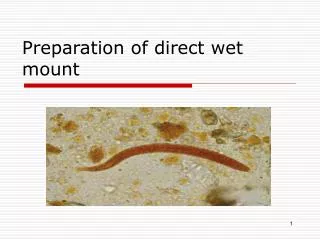
Place discharge on Microscope Slide Place one smear of discharge on one side of the slide and another smear on the other side of the slide. Avoid placing too much discharge on the slide as a thick specimen interferes with readability of the slide. Click her to go back Click here to move on
Add NaCl and KOH Add one drop of NaCL to one area of the discharge and one drop of KOH to the other side and mix with end of wooden stick/swab Observe for a fishy odor with the addition of KOH (whiff test) which is an indicator of bacterial vaginosis (positive whiff test)Two Apply a coverslip to each side The saline side is used to visualize Trichomonads and Clue Cells The KOH side is used to visualize yeast Click her to go back Click here to move on
A fishy odor with the addition of KOH to vaginal discharge is known as ____________ and is indicative of ___________________. A negative whiff test; Bacterial Vaginosis A positive whiff test; Candida A positive whiff test; Bacterial Vaginosis Click here to select this answer Click here to select this answer Click here to select this answer Click her to go back
The correct answer is……………………….. A positive whiff test; Bacterial Vaginosis Click her to go back Click here to move on
Microscopic Examination Begin on low power. Scan the entire slide- observing for yeast and Trichomonads on the saline side and clue cells on the KOH side. Click her to go back Click here to move on
Candida (Yeast) Infection • A diagnosis of Candida vaginitis is suggested clinically by the presence of external dysuria and vulvar pruritis, pain, swelling, and redness. Signs include vulvar edema, fissures, excoriations, or thick curdy vaginal discharge. • The vaginal ph is <4.5 • Wet mount examination may reveal budding yeast and/or pseudohyphae. • Use of 10% KOH in wet preparations improves the visualization of yeast and mycelia by disrupting cellular material that might obscure the yeast or pseudohyphae. Click her to go back Click here to move on
Budding yeasts and Pseudohyphae The morphology is typical of actively growing Candida sp. This slide is also typical of one that has been allowed to ‘incubate’ at room temperature for about 3 hours so that the yeast cells tend to swell. Fresh specimens do not typically exhibit such luxurious growth and vacuolated yeast cells. Budding yeast Pseudohyphae Click her to go back Click here to move on
Budding yeasts and Pseudohyphae Low Power Click her to go back Click here to move on
Click her to go back Click here to move on
Pseudohyphae These are fragile tube-like structures that arise through elongation of the yeast form of Candida. Pseudohyphe may demonstrate a terminal swollen remnant of the original yeast cell. They are called pseudohyphae because they lack true branching as seen with mold like fungi. The side walls are parallel to each other which is an important characteristic that helps separate pseudohyphae from artifact whose side walls vary in width. Small oval structures called bastoconidia are often seen attached along the length of the pseudohyphae. The blastoconidia are smaller in size when compared to the yeast form of Candida. While pseudohyphae are usually seen along with yeast cells or budding yeast, it is also possible to see pseudohyphae in the absence of yeast cells. Pseudohyphae with budding yeast cells Click her to go back Click here to move on
Fragile tube-like structures that arise through elongation of the yeast form of Candida are known as ____________. Budding yeast Trichomonads Clue Cells Pseudohyphae Click here to select this answer Click here to select this answer Click here to select this answer Click here to select this answer Click her to go back
The correct answer is…… Pseudohyphae Click her to go back Click here to move on
Artifact vs. Pseudohyphae One of the more troublesome artifacts is fibers that are sometimes confused with pseudohyphae. There are a few tips that may help in the differentiation. 1. Fibers are generally larger in size that pseudohyphae 2. Pseudohyphae have parallel sides with a consistent dimension between the sides while fibers show variable widths along the fiber. 3. Fibers tend to be birefringent. That is they change color when focusing up and down on the object. Colors are often gold or blue and result from the microscope light being refracted by the fiber. ARTIFACT Click her to go back Click here to move on
Trichomoniasis • Trichomoniasis is caused by the protozoan T. vaginalis. • Many infected women have symptoms characterized by a diffuse, malodorous, yellow-green vaginal discharge with vulvar irritation. However, some women have minimal or no symptoms. • Diagnosis of vaginal trichomoniasis is usually performed by microscopy of vaginal secretions, but this method has a sensitivity of only approximately 60%–70% and requires immediate evaluation of wet preparation slide for optimal results. Click her to go back Click here to move on
Trichomonas • Trichomonas are parasitic protozoa. • They can be very motile • When they begin to die (within 10 minutes of specimen collection), they become sedentary and begin to round up. • Trichomonas should only be reported when motility is observed Click her to go back Click here to move on
True or False: Trichomonas generally begin to lose motility within 20 minutes of specimen collection True False Click here to select this answer Click here to select this answer Click her to go back
The correct answer is…………………. False…….Trichomonas begin to die about 10 minutes after specimen collection. Therefore, it is imperative that the specimen be processed quickly following collection. Click her to go back Click here to move on
Trichomonas Click her to go back Click here to move on
Trichomonas • Trichomonas have a very complex structure. • They have four flagella facing ‘forward’ and a fifth facing ‘backward’ which is attached to an undulating membrane. • The cells are oval in shape, 10-23 µm in length Click her to go back Click here to move on
Trichomoniasis • Appearance of a “strawberry cervix” may be seen with Trichomonas infection Click her to go back Click here to move on
BACTERIAL VAGINOSIS (BV) Must have 3 of the 4 signs for a diagnosis of BV (AMSEL’S CRITERIA) • Discharge, white & homogeneous • Elevated pH, >4.5 • Amine odor present • 20% clue cells present on wet mount Click her to go back Click here to move on
White, homogeneous discharge of BV Click her to go back Click here to move on
Agents of Bacterial Vaginosis • Gardnerellavaginosis(small, gram negative [red] rounded rods - coccobacilli) • Mobiluncus sp. (small, slender gram negative [red] curved rods). • Both are normally found in the vagina (i.e., normal flora) • In bacterial vaginosis, these bacteria increase in number and degrade proteins in the vagina to form the amines that cause the characteristic unpleasant odor associated with BV. Click her to go back Click here to move on
True or False: The vaginal pH is elevated in Bacterial Vaginosis True False Click here to select this answer Click here to select this answer Click her to go back
The correct answer is…………………. True…….The vaginal pH of greater than 4.5 is characteristic of BV. This is an alkaline state. Additional criteria (other than an elevated pH) are needed to make a diagnosis of BV. Click her to go back Click here to move on
Clue cells vs. Normal Squamous Epithelial Cells Clue Cells Squamous Epithelial cells The cell nucleus and the cell boundary are clearly observed. • Clue cells are squamous epithelial cells that are covered with a thick matte of bacterial cells and are associated with bacterial vaginosis. • The traditional definition of a clue cell is that the bacterial overgrowth is so thick that all cellular detail (such as the cell nucleus and the cellular edge) is totally obscured. • It is sometimes possible to detect the nucleus in a clue cell by using the fine focus knob to focus throughout the cell. Click her to go back Click here to move on
Clue cells vs. Clue Cell Squamous Epithelial cells Click her to go back Click here to move on
Low Power Squamous Epithelial cell – not a clue cell Clue cell Click her to go back Click here to move on
Lactobacilli The bacteria shown in this slide are characteristic of lactobacilli, which is normal flora in women following the onset on menses and will persist as normal flora until menopause. Click her to go back Click here to move on
You have reached the end of this program….. Click here to link to CDC free CME/CNE on vaginitis and STDs Start Over Click here to go back and review previous slide Exit Once you are confident that you are comfortable with all the materials presented, proceed to the Vaginal pH and Wet Mount Testing post test at http://www.classmarker.com/professional/ Your username is the first initial of your first name followed by your full last name. Your password is= nwhealth