Download
1 / 9
90 likes | 240 Vues
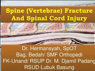
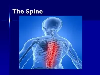
Injury of the spine. Spinal injury carry a double threat: damage to the vertebral column & damage to the neural tissues.

E N D
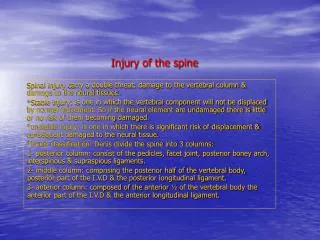
Injury of the spine Spinal injury carry a double threat: damage to the vertebral column & damage to the neural tissues. *Stable injury: is one in which the vertebral component will not be displaced by normal movement. So if the neural element are undamaged there is little or no risk of them becoming damaged. *unstable injury: is one in which there is significant risk of displacement & consequent damaged to the neural tissue. *Denis classification: Denis divide the spine into 3 columns: 1- posterior column: consist of the pedicles, facet joint, posterior boney arch, interspinous & supraspious ligaments. 2- middle column: comprising the posterior half of the vertebral body, posterior part of the I.V.D & the posterior longitudinal ligament. 3- anterior column: composed of the anterior ½ of the vertebral body the anterior part of the I.V.D & the anterior longitudinal ligament.
All #s.that involve the middle column & at least one other column should be regarded as unstable which constitute 10% of all spinal #s. & only 5% associate with cord damage. Mechanism of injury: 1- traction due to resisted muscle effort 2- direct as in penetrating injury. 3- indirect as a result of a fall from a height which may cause flexion, axial compression, flexion distraction, extension, or a combination of forces. *NB. If anterior wedging >40%of vertebral body it will cause progressive flexion deformity “ kyphosis” Diagnosis: 1- history: of trauma above the clavicle, fall from a height, high speed deceleration accident, or post traumatic neck or back pain. 2- examination: a) neck & back for bruises, penetrating injury, deformity, tenderness, or gab. b) shock: there are 3 type of shock may occur in spinal injury 1-hypovolemic 2-neurogenic 3-spinal shock which last for less than 48 hours.
c) neurological examination which include:1- sensory, motor, reflexes. 2- examination for the function of the spinal longitudinal columns: the anterolaterl, posterolateral & posterior column. 3- sacral sparing reflexes: big toe flexion, anal tone, & perianal sensation. 3- imaging: 1- plain x ray: include AP, lateral, open mouth, oblique. 2-CT: show structural damage. 3- CT myelo which is replaced by MRM. 4- MRI. 5- 3D CT or spiral CT specially show odontoid # *Principle of definitive management: objectives: 1- preserve neurological function. 2- relieve any reversible neural compression. 3- restore alignment of the spine. 4- stabilize the spine. 5- rehabilitate the patient.
A) Patient with no neurological injury: 1- if the spine is stable: is by bed rest & support as a collar for the cervical spine & brace for the lumbar spine .there is an exception for the burst # & if the kyphosis deformity is > 30 degrees which need surgical stabilization for better rehabilitation & to prevent progressive deformity. 2- if the spine is unstable: should be held secure until the tissue heal, A)- for the cervical spine is by tongs or halo device, B)- & for the thoracolumbar spine is by internal fixation C)- for dislocation or subluxation is to be reduced by 1- adjusting the posture, 2- traction, 3- open operation. B) Patient with neurological injury: 1- if the spine is stable: rare, is conservative treatment & rehabilitation as soon as possible. 2- if the spine is unstable: a) Conservative in special spine unite where there is 1- round the clock nursing, 2- 2 hourly turning routine, 3- skin toilet, 4- bladder care, 5- specialized physiotherapy, 6- occupational therapy.
b) Operative: open reduction or decompression & stabilization. Benefits are: 1- facilitate nursing. 2- reduce risk of spinal deformity. 3- reduce pain. 4- speeds rehabilitation. BN. i.v methyl pridnisolone has been shown to improve out come if given within 8 hour of injury. Cervical spine: Diagnostic pitfalls in children: 1- atlantodental interval normally <3 mm. but in children it reach up to 4.5 mm. because the skeleton incompletely ossified& the ligaments are relatively lax. 2- apparent subluxation of C2/C3 pseudosubluxation. 3- retropharyngeal space normally 5mm. Above the trachea, & < than 1 vertebral width below it increase in children by forced expiration during crying. 4- growth plates may be mistaken for #. 1)- at the base of the odontoid fuse by the age of years 2)- at the base of the spinous processes. 3)- at the tip of the odontoid. 5- SCIWORA = SPINAL CORD INJURY WITHOUT RADIOGRAPHIC ABNORMALITY. Normal radiograph in children not exclude spinal cord injury.
C1 # Jefferson’s #:#of the ring of the atlas caused by sudden sever load on the top of the head, there is no encroachment on the canal & usually no neurological damage X- ray: open mouth view show separation of the lateral mass away from the odontoid pig CT. better define the #. if undisplaced it is usually stable & treated by semi rigid collar or hallo vest, but if displaced > 7 mm. usually unstable & treated by hallo vest or C1/C2 arthrodesis. Odontoid # of C2: caused in young adult by flexion injury occur in high velocity accident or sever fall, while in elderly osteoporotic people occur as a result of hyperextension injury caused by fall on to the face. Neurological damage occur in 25% of cases. X- ray: show 3 type of # 1)- avulsion # of the tip of the odontoid usually stable. 2)- # at the junction of the base of the odontoid & the body of the axis usually unstable 3)- # through the body of the axis which is usually stable. Rx. Type 1 immobilization in semi rigid collar until discomfort subside.
Type 2: a) undisplaced treated by halo vest. b) displaced > 5 mm. reduced by traction then operative screw fixation or halo vest. Type 3: a) undisplaced halo vest. b) if displaced reduction by traction then halo vest for 8-12 wk. # C2 pedicles “Hangman #” : # of the C2 pedicles with rupture of C2/C3 disc caused by extension & distraction force, the # usually unstable. Rx. : if the # undisplaced which is usually stable halo vest immobilization. If displaced which is usually unstable then reduction but traction should be avoided here “because the mechanism of injury is distraction” then halo vest. For persistent instability or when associated with C2/C3 facet dislocation then open reduction & stabilization required. Tear drop #: # of the anteroinferior part of the vertebral body usually caused by axial compression & flexion force, it is very unstable injury usually associated with middle column & posterior element damage, so treated by anterior or posterior stabilization & if there is sing of cord impingement so decompression also required.
Sprained neck “ whiplash injury ”soft tissue sprain or wrenching injury of the neck occur in rear-end collision in which the body is thrown forward & the head jerked backward with hyper extension of the lower cervical spine there is strain of the anterior longitudinal ligament, the capsular fiber of the facet joint, some time there is also damage to the I.V.D. C/F: after 12-48 hour of accident patient develop neck pain, stiffness & tenderness. Some time associated with headache, dizziness, blurring of vision, parasthesia in the arms, T.M joint discomfort & tinnitus. X-ray: show straightening of the otherwise normal cervical lordosis, “a sing of muscle spasm” D.Dx.: other cause of neck pain & stiffness: 1-# of the cervical spine. 2- I.V.D herniation 3- seat belt injury cause traction on the supra scapular nerve or brachial plexus give symptom like whiplash injury. Rx.: analgesic medication in the first few Wks. Then graded exercises encouraged. Thoracolumbar & lumbar injuries: Burst#: the vertebral body exploded & the posterior part of the vertebral body is shattered & fragments of bone & disc may be displaced into the spinal canal, both the anterior & the middle column are failed & the # is usually unstable. It is caused by sever axial compression. X-ray: show increase inter pedicular distance & retro pulsed body fragment better seen by C.T
Rx.: 1- if there is minimal retropulsion of bone, no neurological damage & minimal anterior wedging, patient kept in bed until acute symptoms subside then mobilized in thoracolumbar brace for 12 Wk. 2- if patient develop sign of neurological damage this call for anterior decompression & stabilization Jack-knife injury: Typically occur in lap seatbelt injury when the body is thrown forward against the restraining strap resulting in flexion & distraction causing the mid lumbar spine to jack- knife around an axis placed anterior to the vertebral column. The posterior & middle columns fail in distraction, & the # unstable in flexion. The tear pass transverse through the bone “Chance #” or through the ligaments, or both Rx.: Chance # being all boney heal rapidly & require no more than 3 month in plaster jacket, ligamentous injury usually unstable & require posterior spinal fusion.