Download
1 / 54
550 likes | 683 Vues
Pathophysiology Treatments. Main damages in the brain of neonates born prematurely or at term. The more fragile infants are those born before 30 weeks : Important developmental aspects in structures and metabolism. Neuronal migration, blood brain barrier, cells ’ biochemistry,

E N D
Pathophysiology Treatments Main damages in the brainof neonatesbornprematurely or atterm Pr O Battisti, brain damage in neonates
The more fragile infants are thosebornbefore 30 weeks:Important developmental aspects in structures and metabolism Neuronal migration, blood brain barrier, cells ’ biochemistry, Cerebral blood flow autoregulation 2 Pr O Battisti, brain damage in neonates
Anatomic aspectsThe quantitative brain in newborns Brain is 14-16 % of body weight Brain is 60 % of body metabolism ( see glucose ) and 02 consumption 10 % of brain is CSF of which 6/7 is coming from choroid plexus and 1/7 from capillaries 3-5 % of brain is blood 40 % of brain is glial cells 4% of brain is neurons 40% of brain is EC fluid 5 Pr O Battisti, brain damage in neonates
Fetal and neonatalBraindevelopment: histology and biochemistry Microcirculation: observe differences between A and V • Radial cells; • oligodendrocytes • astrocytes • microglia 40 % glial cells , 4 % neurons, 4 % blood, 10 % CSF, 35 % ECF, 10 % variance 6 Pr O Battisti, brain damage in neonates
The late neuronal migration (Sarnat and Sarnat) At 28 weeks At 24 weeks 7 Pr O Battisti, brain damage in neonates
iCDG Extrinsic inflammation In 25 – 33 % cases 8 Pr O Battisti, brain damage in neonates
The concept of neurone-glial cells association Oligodendrocytes: Perineuronal; Astrocytes: perivascular, in White and grey matter Radial cells Microcytes: travellers Layers 1, 2 and 3 for intra-cortical associative relationships 1, 4, 5 , 6 for projective intra-cortical and subcortical relationships Pr O Battisti, brain damage in neonates
4 % 6 % 20 % % 40 % 50 % 80 % Pr O Battisti, brain damage in neonates
Summary of Main features concerning brain cytology and biochemistry Function and integrity of cells, temperature; 10 glial cells for one neuron; Neurone-glial cells unit; Surrounding capillaries and the BBB; Differences in density of capillaries; Differences in veins and arteries networks; Differences in CMRO2 and CMRG between neurons and glial cells Late neuronal migration and transitory period of hypersensitivity - Neurons: similarity of reactions -Oligodendrocytes: oxydative stress protection ; trophins; myelin synthesis; non phagocytic - Radial cells: guiders and helpers - Astrocytes: nutrition of neurons, reservoir of beta-amyloid protein and chondroitine sulfate proteoglycan glutamate and TNF;« fibrous » in WM, « protoplasmic » in GM; - Microglia ( < mesoderm ): macrophage, reservoir of cytokins 11 Pr O Battisti, brain damage in neonates
Metabolic aspectsIntracerebralConsequences of iCDO2 and/or iCDG At BBB level: entry of small molecules (15 ‘ ) and big molecules ( 120 ‘); of neutrophils and monocytes ( 120 ‘ ) At neurons level: axones then dendrites edema ( 30-55 ‘ ), followed by retraction and hypersensitivity to EAA; body edema ( 50-75 ‘), action on peptides and nucleus; At vessels’level :capillaries surrounded, , thrombosis; At OL level: At astrocytes level: glutamate, NO, FR, proteolytic enzymes At microglia level: Energy failure and oxydative stress: Free radicals ( OFR, NO, Fe+++ ) EAA Release of NA from locus ceorulus Activation of microglia and LycT4 Genes activation ( CREB, JUN Relesa of toxins: AOAA, MPP, 3NPA Inflammatory products: - Proteolytic enzymes on matrix: from neurons, astrocytes, microglia; - Il 1,6,8,9, TNFa, complement, antithrombin III, factor V, protein C, antipohospolipid antibody 12 Pr O Battisti, brain damage in neonates
Excitotoxicity: apoptosis and necrosis;neuroplasticity Prof Oreste Battisti 13 Pr O Battisti, brain damage in neonates
Cerebral blood flow and metabolic autoregulation loss or absence • CBF absent if: - respiratory distress; - circulatory distress; - hypoglycemia; - CNS infection; - brain trauma; • Loosing the independence • 1° Systolic blood pressure; 2 °CO2 pressure (attention to pCO2 < 27 for >= 1hr or > 65 for > 6 hrs ); 3° O2 content; Locus ceroelus Pr O Battisti, brain damage in neonates
Cbf ml/100g/m [ aG ] ->I [ aG ]-> W [ aO2]->I [aO2]-> W 20 21 36 10 22 15 30 50 13 29# 10 40 72 19 44# 2 82 143 28# 62# Glucose ( mg/dL ) and O2 ( ml/dL ) requirements in the distressed brain Hemoglobin from present blood stores O2 for 60 sec Hb 12 g/dL term Preterm « good » target Preterm sick Hb 15 g/dL Glycogen stores are enough for 30-60 minutes... Pr O Battisti, brain damage in neonates
The functional unit in the brain: a close relationship between cells and vessels Pr O Battisti, brain damage in neonates
Cell temperature, brain perfusion, function, integrity and metabolism Prof Oreste Battisti 18 Pr O Battisti, brain damage in neonates
Disturbances of late neuronal migration: axonal retraction, pericapillaries congruence, rosettes, ependymal dysruption, And also of volume and Myelination 19 Pr O Battisti, brain damage in neonates
From protecting to damaging « biochemical attitudes » Depending on the environmental conditions, actual and preceding, factors can protect or damage iCe and iNu Ca++ FR CREB, CAMD, caspase 3 and 6 • Appoptotic cascade : • - if > 1h ofLow pCO2 ( < 27 mmHg; ) • - if > 6 h of high pCO2 ( > 65 mmHg ) • if hypoxia ( > 65 mmHg? pO2 ) • for > 1 h ? Time ? 20 Pr O Battisti, brain damage in neonates
damagerepair Similarity of reactions by neurons; more dependency of environment damage from inside CNS: --> astrocytes, loss of nutrition, of cytoskeletal compound, activation of GAP3 and release of toxins --> microglia, release of toxins and activation of MHCII and CR3 --> neurons: loss of targed-derived factors; dendritic and axons atrophy and loss of molecules transport --> low synthesis of growth factors and low leullar guidance impairement of BBB and arrival of cytokines ( lungs, digestive tract, blood cells, bone marrow ) Neurotrophic factors have specific targets; their main roles are to prevent neuronal loss and maintenance of axons regrowth; 1.Neurotrophins family (NGF, BDNF ) 2.Cytokins growth factors ( LIF, CNTF, CT-1 ) 3. Fibroblats growth factors ( FGF1 and 2 ); mainly for layers II and III, hippocampus. 4. The insulin growth factors 5. The transforming factor beta family of growth factors; 6. Epidermal growth factor 7.Hepatocyte GF and Immunophilins 21 Pr O Battisti, brain damage in neonates
Endotoxins coming from outside brain affects heart Function and possibly CBF = « dual effects » Or Inflammation 22 Pr O Battisti, brain damage in neonates
The cellular effects of hypoxia or iCDO2 In about 75 % of cases, first trigger is iCDO2 giving rise to excitotoxycity and ifnflammatory cascade; In about 25 % of cases, first trigger is inflammation cascade The importance of iCDG per se is qualitatively well known, but its confounded and proportional place can’t be quantified EAA, Free radicals and cytokins 23 Pr O Battisti, brain damage in neonates
Ultrastructure of blood-brain-barrier:this very important structure is not efficient before 27 weeks, and definitely not before 32 weeks 24 Pr O Battisti, brain damage in neonates
The blood-brain barrier Brain cells: neurons, astrocytes, microcytes,radial cells, oligodendrocytes; Microcirculation; Ependyma and villi; Arachnoids; 25 Pr O Battisti, brain damage in neonates
This is about in 70- 80 % of cases This is in about 15- 25 % of cases - Late migration - CBF autoreg 26 Pr O Battisti, brain damage in neonates
Cytokines liberation and storage Density of small vessels in germinal > cortex > white matter; Capillaries in BBB are very rich in mitochondria; All Brain cells can produce complement, cytokines and coagulation proteins; MHC system in brain = HLA system outside brain; Microglia works with CD4+ TH1 cells; can command astrocytes and endothelial cells. Astrocytes can store toxins Ly B works with CD4+TH2 cells; Any cell works with CD8+T cells Fibroblasts can produce cytokines and stem cells. Distant intervention By locus coeruleus And hippocampus 27 Pr O Battisti, brain damage in neonates
factors 28 Pr O Battisti, brain damage in neonates
Some important « apples » Pathology of leucomalacia: « end product » Early and late neuronal migrations: their importance Cerebral blood flow autoregulation impairement Biochemical properties of brain cells Metabolic differences between neurons and glial cells pH, pCO2, BP, O2 intervention Cerebral blood flow, CMRG, CMRO2 and extractions measurements US diagnosis of PVL and classification Epidemiological studies The inflammatory components 29 Pr O Battisti, brain damage in neonates
neuroprotection: why hypothermy will not be possible in premature infants Values at 27 ° C ! 30 Pr O Battisti, brain damage in neonates
The GLUT or glucose transporters Pr O Battisti, brain damage in neonates
Central role of glucose for energy an synthesis • ATP and 5-Pentose; • In- and out-cells composition • neurotransmission • Defenses against FR and EAA • BMR • Muscles ( FFA ) and intestines ( glutamine and KB ) have alternatives Pr O Battisti, brain damage in neonates
Neurones and glial ( ) survival times in secaccording to CBF in ml/100g/min, [O2]a in ml/dL= 1.34 Hbg/dL * Sat + (0.0224 pO2 mmHg)coefficient of extraction maximal =0.75 Prof Oreste Battisti, CHU-NDB, ULG
Glucose and energy in cells: cytoplasm and mitochondriaalso dependent of body temperature > 95 % In normal cell: G + O2 Free radicals ATP + CO2 + H2O coupled radicals < 5% In cellwith mitochondrialimpairement: G + O2 hypoxia reoxygenation ATP + CO2 + lactic acid Loss of energy and Electrolytes disturbances Free radicals Pr O Battisti, brain damage in neonates
Neonatal resuscitation with room air vs oxygen From Saugstad, animal&clinical data Prof Oreste Battisti, CHU-NDB, ULG
The intertissular metabolic integration 15 % body mass; 60 % BMR • About nutritional needs and storages: -> aminoacids, fat and Carbohydrates; -> flow autoregulation; • About cerebral growth: - in preterms; - in case of retarded growth; • About muscles growth: - in preterms; - in case of retarded growth; muscles 24 % body mass Pr O Battisti, brain damage in neonates
Kcal/kg/d QO2 l/kg/m 120 6-8 120 6-8 % Pt 20-30 10-15 % Lp < 5 25-30 % Gl 60-70 50-60 The last 3 weeks of pregnancy prepares the nervous and endocrine systems to neonatal adaptation, mainly the autonomic nervous system Pr O Battisti, brain damage in neonates
About the periods of sleep • REM-periods:under adrenergic control; synthesis of proteins and neurotransmitters; high metabolic rate. Correlation with growth. • Non REM-periods: under vagal control; pauses in synthesis; lower metabolic rate. Pr O Battisti, brain damage in neonates
The more fragile infants are thosebornbefore 30 weeks:Important developmental aspects in structures and metabolism Neuronal migration, blood brain barrier, cells ’ biochemistry, Cerebral blood flow autoregulation 40 Pr O Battisti, brain damage in neonates
(Paneth ) SUMMARY OF 5 KEY FINDINGS ABOUT PREMATURE INFANTS IN DEN AND NBH Brain damage is widespread in infants who die, and can be diffuse or focal, but white matter is the tissue most affected. US evidence of white matter injury ... is the most important determinant of long-term outcome. Thyroid hormone is the single most predictive measure of outcome obtainable on serum in the first week of life Hypocapnia (PCO2 < 25) and perhaps hyperoxia (PO2 > 60) should be avoided. The finding in the placenta most predictive of brain injury is fetal vasculitis; membrane inflammation alone is not associated with brain injury • Comment: • Amnionitis gives: • an increased release of cytokines and • a decreased expression of angiogenic factors 41 Pr O Battisti, brain damage in neonates
Facteurs à considerer • La chorioamniotite • Le RCIU • La grande prématurité - La détresse respiratoire, la dysplasie BP, hypothyroïdie, hyperbilirubinémie, les perturbations hémodynamiques • L’entérocolite nécrosante Pr O Battisti, brain damage in neonates
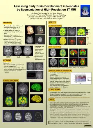
Studying Brain Development 9 months 10 weeks premature Term Pr O Battisti, brain damage in neonates
Effet structural de l ’hypoxie sur les cellules cérébrales Prof Oreste Battisti, CHU-NDB, ULG
Disturbances of late neuronal migration: axonal retraction, pericapillaries congruence, rosettes, ependymal dysruption, And also of volume and Myelination 48 Pr O Battisti, brain damage in neonates
Voxel Labeling:Do you see the tracts Cingulum Cingulum, arcuate fasciculus, uncinate fasciculus Pr O Battisti, brain damage in neonates