Download
1 / 70
720 likes | 730 Vues
Genes and Genetic Diseases. Chapter 2. DNA. Pentose sugar (deoxyribose) Phosphate molecule Four nitrogenous bases: Pyrimidines: cytosine and thymine Purines: adenine and guanine Double helix model Nucleotide. DNA (cont’d). Proteins. One or more polypeptides Composed of amino acids

E N D
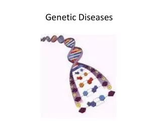
Genes and Genetic Diseases Chapter 2
DNA Pentose sugar (deoxyribose) Phosphate molecule Four nitrogenous bases: Pyrimidines: cytosine and thymine Purines: adenine and guanine Double helix model Nucleotide
Proteins One or more polypeptides Composed of amino acids Twenty amino acids Directed by sequence of bases (codons)
DNA Replication Untwisting and unzipping of the DNA strand Single strand acts as a template Complementary base pairing by DNA polymerase Adenine-thymine; cytosine-guanine
Mutation Any inherited alteration of genetic material Chromosome-aberrations Base pair substitution One base pair is substituted for another Frameshift mutation Insertion or deletion of one or more base pairs Causes a change in the entire “reading frame”
Mutation (cont’d) Spontaneous mutation Mutation that occurs in absence of exposure to known mutagens Mutational hot spots Areas of the chromosomes that have high mutation rates A cytosine base followed by a guanine is known to account for a disproportionately large percentage of disease-causing mutations
Mutagen Agent known to increase the frequency of mutations Radiation Chemicals Nitrogen mustard, vinyl chloride, alkylating agents, formaldehyde, sodium nitrite
Transcription RNA is synthesized from the DNA template RNA polymerase binds to promoter site Results in the formation of messenger RNA (mRNA) RNA polymerase detaches mRNA moves out of the nucleus and into the cytoplasm Transcription continues until termination sequence is reached
Gene Splicing Many RNA sequences are removed, (introns) and remaining are spliced together (exons) before mRNA migrates to the cytoplasm
Translation Process by which RNA directs the synthesis of a polypeptide via interaction with tRNA Site of protein synthesis is the ribosome tRNA contains a sequence of nucleotides (anticodon) complementary to the triad of nucleotides on the mRNA strand (codon)
Translation (cont’d) The ribosome moves along the mRNA sequence to translate the amino acid sequence
Chromosomes Somatic cells: Contain 46 chromosomes (23 pairs) Diploid cells Gametes: Contain 23 chromosomes Haploid cells One member of each chromosome pair
Chromosomes (cont’d) Meiosis Formation of haploid cells from diploid cells Mitosis Formation of somatic cells
Chromosomes (cont’d) Autosomes The first 22 of the 23 pairs of chromosomes in males and females The two members are virtually identical and thus said to be homologous Sex chromosomes Remaining pair of chromosomes In females, it is a homologous pair (XX) In males, it is a nonhomologous pair (XY) Karyotype (karyogram)
Karyotype Ordered display of chromosomes
Chromosome Aberrations Euploid cells have a multiple of the normal number of chromosomes Haploid and diploid cells are euploid forms When a euploid cell has more than the diploid number, it is called a polyploid cell Triploidy: a zygote having three copies of each chromosome (69) Tetraploidy: four copies of each (92 total) Neither triploid nor tetraploid fetuses survive
Chromosome Aberrations (cont’d) Aneuploidy A somatic cell that does not contain a multiple of 23 chromosomes A cell containing three copies of one chromosome is trisomic (trisomy) Monosomy is the presence of only one copy of any chromosome Monosomy is often lethal, but infants can survive with trisomy of certain chromosomes “It is better to have extra than less”
Chromosome Aberrations (cont’d) Disjunction Normal separation of chromosomes during cell division Nondisjunction Usually the cause of aneuploidy Failure of homologous chromosomes or sister chromatids to separate normally during meiosis or mitosis
Autosomal Aneuploidy Partial trisomy Only an extra portion of a chromosome is present in each cell Chromosomal mosaics Trisomies occurring only in some cells of the body
Autosomal Aneuploidy (cont’d) Down syndrome Best known example of aneuploidy Trisomy 21 1:800 live births Mentally retarded, low nasal bridge, epicanthal folds, protruding tongue, poor muscle tone Risk increases with maternal age >35
Sex Chromosome Aneuploidy One of the most common is trisomy X (a female that has three X chromosomes) Symptoms are variable: sterility, menstrual irregularity, and/or mental retardation Symptoms worsen with each additional X
Sex Chromosome Aneuploidy (cont’d) Turner syndrome Females with only one X chromosome Characteristics: Underdeveloped ovaries (sterile) Short stature (~ 4'7") Webbing of the neck Edema Underdeveloped breasts; wide nipples High number of aborted fetuses X is usually inherited from mother
Sex Chromosome Aneuploidy Klinefelter syndrome Individuals with at least two Xs and one Y chromosome Characteristics Male appearance Develop female-like breasts Small testes Sparse body hair Long limbs
Sex Chromosome Aneuploidy (cont’d) Klinefelter syndrome (cont’d) Some individuals can be XXY and XXXY The abnormalities increase with each X
Abnormalities in Chromosome Structure Chromosome breakage If a chromosome break does occur, physiologic mechanisms will usually repair the break, but the breaks often heal in a way that alters the structure of the chromosome Clastogens Ionizing radiation, chemicals, and viruses
Abnormalities in Chromosome Structure (cont’d) Breakage or loss of DNA (deletions) Cri du chat syndrome “Cry of the cat” Deletion of short arm of chromosome 5 Low birth weight, metal retardation, and microcephaly
Abnormalities in Chromosome Structure (cont’d) Duplication Presence of a repeated gene or gene sequence Less serious consequences because better to have more genetic material than less (deletion) Duplication in the same region as cri du chat causes mental retardation but no physical abnormalities
Alterations in Chromosome Structure Inversions Two breaks on a chromosome Reversal of the gene order Usually occurs from a breakage that gets reversed during reattachment ABCDEFG may become ABEDCFG Position effect
Abnormalities in Chromosome Structure Translocations The interchanging of material between nonhomologous chromosomes Reciprocal translocation occurs when two chromosomes break and the segments are rejoined in an abnormal arrangement Robertsonian translocation occurs when fusion at centromere, forming a single chromosome
Abnormalities in Chromosome Structure (cont’d) Fragile sites Fragile sites are areas on chromosomes that develop distinctive breaks or gaps when cells are cultured No apparent relationship to disease
Abnormalities in Chromosome Structure (cont’d) Fragile X syndrome Site on the long arm of the X chromosome Associated with mental retardation; second in occurrence to Down syndrome Higher incidence in males because they have only one X chromosome
Genetics Locus Position of a gene along a chromosome Allele A different form of a particular gene at a given locus Example: Hgb A vs. Hgb S Polymorphism Locus that has two or more alleles that occur with appreciable frequency
Genetics (cont’d) Homozygous Loci on a pair of chromosomes have identical genes Example O blood type (OO) Heterozygous Loci on a pair of chromosomes have different genes Example AB blood type (A and B genes on pair of loci)
Genetics (cont’d) Genotype (“what they have”) The genetic makeup of an organism Phenotype (“what they demonstrate”) The observable, detectable, or outward appearance of the genetics of an organism Example A person with the A blood type could be AA or AO: A is the phenotype; AA or AO is the genotype
Genetics (cont’d) If two alleles are found together, the allele that is observable is dominant, and the one whose effects are hidden is recessive In genetics, the dominant allele is represented by a capital letter, and the recessive by a lowercase letter Alleles can be codominant
Genetics (cont’d) Carrier A carrier is one who has a disease gene but is phenotypically normal For a person to demonstrate a recessive disease, the pair of recessive genes must be inherited Example Ss = sickle cell anemia carrier ss = demonstrates sickle cell disease
Genetics (cont’d) Transmission of genetic disease Mode of inheritance Principle of segregation Principle of individual assortment Chromosome theory of inheritance
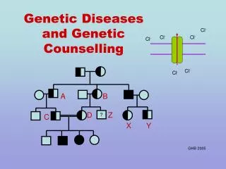
Pedigrees Used to study specific genetic disorders within families Begins with the proband
Single-Gene Disorders Autosomal dominant disorder Abnormal allele is dominant, normal allele is recessive, and the genes exist on a pair of autosomes