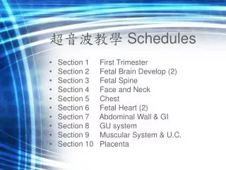
超音波教學 Schedules
超音波教學 Schedules. Section 1 First Trimester Section 2 Fetal Brain Develop (2) Section 3 Fetal Spine Section 4 Face and Neck Section 5 Chest Section 6 Fetal Heart (2) Section 7 Abdominal Wall & GI Section 8 GU system Section 9 Muscular System & U.C.

超音波教學 Schedules
E N D
Presentation Transcript
超音波教學 Schedules • Section 1 First Trimester • Section 2 Fetal Brain Develop (2) • Section 3 Fetal Spine • Section 4 Face and Neck • Section 5 Chest • Section 6 Fetal Heart (2) • Section 7 Abdominal Wall & GI • Section 8 GU system • Section 9 Muscular System & U.C. • Section 10 Placenta
超音波教學Section I • Speaker • 陳志堯醫師 • Director • 趙灌中主任 洪正修主任 • August 25, 2006
Topic 1 Normal Early Pregnancy
Day 13 Day 21 Day 28
AL, allantois; CF, chorion frondosum; CL, chorion laeve; CS, connecting stalk; DV, decidua vera (along endometrium; E, embryo; ECS, extrachorionic space (endometrial cavity); EEC, extraembryonic coelom; EEM, extraembryonic mesoderm; OM, omphalomesenteric duct; PG, primary gut, Y1 primary yolk sac; Y2, secondary yolk sac.
Blastocyst implantation site in spontaneous pregnancies 89.1% had E-GSs detected in the upper region, which was found to be the most frequent region. Minami S, J Nippon Med Sch. 2003 Jun;70(3):250-4.
Anembryonic Pregnancy • Mean sac diameter > 10mm with no Y.S. • Mean sac diameter > 18mm with no Emb. • Empty amnion. • Poor color signal around sac.
Potential Pitfalls • Mistaking yolk sac for embryo
Potential Pitfalls • Mistaking Y.S. for embryo • Mistaking sub-chorionic hemorrhage for G.S.
Potential Pitfalls • Mistaking Y.S. for embryo • Mistaking sub-chorionic hemorrhage for G.S. • Missing multiple G.S.
IUP Lt adnexal ectopic sac
Potential Pitfalls • Mistaking Y.S. for embryo • Mistaking sub-chorionic hemorrhage for G.S. • Missing multiple G.S. • Pseudo-sac of E.P.
Potential Pitfalls • Mistaking Y.S. for embryo • Mistaking sub-chorionic hemorrhage for G.S. • Missing multiple G.S. • Pseudo-sac of E.P. • Normal rhombencephalon for cystic brain mass
Potential Pitfalls • Mistaking Y.S. for embryo • Mistaking sub-chorionic hemorrhage for G.S. • Missing multiple G.S. • Pseudo-sac of E.P. • Normal rhombencephalon for cystic brain mass • Physiological bowel herniation for omphalocele.
Topic 2: 1st Trimester Ultrasound Screening 11~13+6 weeks scan
隋·巢原方在《诸病源候论》 • “妊娠三月名始胎,当此之时,血不流行,形象始化,未有定仪,因感而变。”
妇女怀孕3个月时,胚胎已渐次发育成胎儿,这时虽然已从形体上初步成形。但是还没有定型,即所谓“未有定仪”,其可塑性很大,当孕妇见到带有什么特征的东西,她所怀胎儿的形象,包括胎儿的形体和精神,也就会随之产生变化,这就是“见物而变”的本意。故此给那些想生一个长相漂亮、体力过人、才华出众的孩子的孕妇提出了要求,在孕期内要多想好事,多多行善,多看美好的事物,以期感化腹内的胎儿。通过母亲美与善的良好“见物”刺激,而使胎儿向更加聪明、善良、健康、漂亮的方面“变化”。妇女怀孕3个月时,胚胎已渐次发育成胎儿,这时虽然已从形体上初步成形。但是还没有定型,即所谓“未有定仪”,其可塑性很大,当孕妇见到带有什么特征的东西,她所怀胎儿的形象,包括胎儿的形体和精神,也就会随之产生变化,这就是“见物而变”的本意。故此给那些想生一个长相漂亮、体力过人、才华出众的孩子的孕妇提出了要求,在孕期内要多想好事,多多行善,多看美好的事物,以期感化腹内的胎儿。通过母亲美与善的良好“见物”刺激,而使胎儿向更加聪明、善良、健康、漂亮的方面“变化”。
五帝时帝妃子姜源氏妊娠期间性情清静,喜好稼樯,经常随夫郊祭。观察植物生长,所以她的儿子后稷能种五谷,成为我国农业的第二始祖。这里边即寓有“见物而变”的含义。古人“见物而变”胎教理论的提出,当然受当时文化背景的影响,有时代的局限性,但还是有一定的实际指导意义。五帝时帝妃子姜源氏妊娠期间性情清静,喜好稼樯,经常随夫郊祭。观察植物生长,所以她的儿子后稷能种五谷,成为我国农业的第二始祖。这里边即寓有“见物而变”的含义。古人“见物而变”胎教理论的提出,当然受当时文化背景的影响,有时代的局限性,但还是有一定的实际指导意义。
胎兒NT增厚之病理生理學 • 心臟功能失調 • 頭部及頸部靜脈充血 • 細胞外間質成分轉變 • 淋巴管排泄失能 • 貧血 • 低蛋白血症 • 先天性感染
R. BECKER Ultrasound Obstet Gynecol 2006; 27: 613–618
叫我第一名 6.5~7.4 Karl Oliver Kagan,Obstet Gynecol. 2006 Jan;107(1):6-10.
WHY AND FOR WHOM IN THE FIRSTTRIMESTER? • Previous family history of cardiac defects • The finding of an increased NT at 11 to 14 weeks.
The gestation should be 11-13+6 weeks and the fetal CRL should be 45-84 mm. • In chromosomally normal fetuses the incidence of absent nasal bone is < 1% in Caucasian populations and about 10% in Afro-Caribbeans. • The nasal bone is absent in 65-70% of trisomy 21 fetuses and in > 50% of trisomy 18 and 30% of trisomy 13 fetuses. • For a false positive rate of 5%, screening by a combination of sonography for fetal NT and nasal bone and maternal serum free β-hCG and PAPP-A can potentially identify more than 95% of trisomy 21 pregnancies. Fetal Medicine Foundation, London 2006
Ductus Venosus • At 11-13+6 weeks abnormal ductal flow is observed in 5% chromosomally normal fetuses, in about 80% with trisomy 21 and 75% with all chromosomal abnormalities. • Assessment of the ductus venosus can be combined with measurement of fetal NT to improve the effectiveness of early sonographic screening for trisomy 21