Download
1 / 1
10 likes | 127 Vues
Myeloid Cell Activation in Sickle Cell Disease: The Role of Dysregulated Sphingolipid Metabolism in the Disease State. 1 Lane AR, 2 Awojoodu AO, 2 Botchwey EA 1 School of Biology, Georgia Institute of Technology, Atlanta, GA

E N D
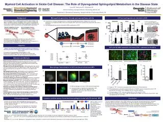
Myeloid Cell Activation in Sickle Cell Disease: The Role of Dysregulated Sphingolipid Metabolism in the Disease State 1Lane AR,2Awojoodu AO, 2Botchwey EA 1School of Biology, Georgia Institute of Technology, Atlanta, GA 2Department of Biomedical Engineering, Georgia Institute of Technology, Atlanta, GA S1P and sphingosine are elevated in SCD Background Microparticle generation through sphingomyelinase activity Sickle cell disease (SCD) is a blood disorder caused by a point mutation in the gene for hemoglobin. Defective hemoglobin results in sickling of red blood cells (RBCs) and increased clotting. Macrophages and monocytes, myeloid-derived immune cells, can adhere at these clots, further contributing to inflammation and vaso-occlusion, which in extreme cases can ultimately result in organ failure1. Sphingolipids are a family of lipids that play a key role in cell membrane dynamics and signaling. Sphingosine 1-phosphate (S1P) is an immunomodulatory sphingolipid that is of special interest for SCD because it is highly concentrated in red blood cells and is elevated in SCD2. Conversion of membrane sphingomyelin to ceramide by acid sphingomyelinase (SMase) can result in the production of microparticles (MPs), which are elevated in SCD, that can be secreted and interact with other cells in circulation. Acid sphingomyelinase activity is elevated in RBCs and plasma in SCD. A) S1P and sphingosine concentrations in AA and SS whole blood, plasma, and RBC. B) S1P and sphingosine concentrations and ratio of ceramide:sphingomyelin in AA and SS microparticles A SS RBC AA RBC Normal hemoglobin β-chain: Val-his-leu-thr-pro-glu-glu- SCD hemoglobin β-chain: Val-his-leu-thr-pro-val-glu A 20 µm Adherent macrophage or monocyte B sickled RBC normal RBC AA MP SS MP Objective B S1P and SS RBC treatment increases THP-1 adhesion to HUVECs * • Explore how dysregulated sphingolipid metabolism contributes to monocyte activation and vascular inflammation in sickle cell disease. Specifically: • Characterize the dysregulation of sphingolipids in SCD • Examine the contribution of S1P to the adhesion of myeloid cells • Examine the effects of RBC-derived microparticles on myeloid cells * * A B Smase activity (nmol/min/mL) * DAPI THP-1 HUVEC THP-1:HUVEC Materials and Methods Non-SCD SCD Non-SCD SCD RBC Plasma RBC Blood collection and harvest. Blood was collected from SCD or non-SCD donors through the Sickle Foundation of Georgia during homeostasis. Blood was fractionated into plasma, peripheral blood mononuclear cells, and RBC using a Ficoll density gradient. Microparticle harvest and quantification. Ultracentrifugation was used to harvest microparticles (MPs) from RBC fractions. MPs were characterized with flow cytometry by staining with FITC-Annexin IV and labeled with CFSE. Sphingolipid quantification.Sphingolipids were extracted with organic solvents before being quantified on a tandem HPLC-MS machine. Macrophage differentiation and polarization. THP-1 monocytes were differentiated to M0 macrophages with PMA and polarized to M1 or M2 macrophages with LPS + IFN-γ or IL-4 respectively. Fluorescent staining of polarized macrophages. Polarized macrophages were stained with DAPI (a nuclear stain) and phalloidin (an actin stain). Monocyte adhesion to HUVEC. THP-1 monocytes were coincubated with confluent HUVECs. Cells were treated with 1 μm S1P or AA/SS plasma for 1 hour or AA/SS RBC for 18 hours and coincubated with HUVEC for 4 hours. Cell counts were taken manually and the ratio of THP-1:HUVEC was calculated, or cells were quantified by DRAQ5 fluorescence. Polarized macrophage internalization of SS-derived CFSE stained microparticles. Polarized macrophages were incubated with MPs for 1, 2, or 4 hours, followed by permeabilization and staining with DAPI and phalloidin. Luminex cytokine assay of polarized macrophages treated with SS-derived MPs. Macrophages were coincubated with MPs for 1, 2, or 4 hours. Cytokines were detected using Luminex cytokine assay. D Non-SCD SCD C Macrophage internalization of SCD red blood cell-derived MPs Non-SCD A SCD B M0 macrophages internalize CFSE-stained microparticles Confocal images of internalized microparticles in M0 macrophages. Intact MPs were observed in the periplasm. In some cases, the CFSE stain filled the periplasm of the cells. Images of THP-1 monocytes coincubated with HUVECs with S1P pretreatment. B) S1P pretreatment of monocytes increased adhesion to HUVECs over 5x compared to control. S1P pretreatment of HUVECs had a negligible effect on monocyte adhesion. Confocal images of THP-1 cells coincubated with non-SCD and SCD RBCs. The THP-1:HUVEC ratio for THP-1 cells coincubated with SS RBCs was almost 4x that of THP-1 cells coincubated with non-SCD RBCs. 0.2 µm Acknowledgements Luminex cytokine assay of polarized macrophages treated with SS-derived MPs Conclusion This work supports a pathway in which RBC sicklingin SCD activates acid sphingomyelinase, altering the distribution and concentration of sphingolipids in the RBC membrane and producing sphingolipid-rich microparticles that are secreted and can interact with cells in circulation, contributing to monocyte activation and vascular inflammation. Pharmacological therapies acting on this pathway may reduce myeloid cell activation, recruitment, and adherence and represent a more effective treatment for this disease. Funding sources include: Petit Undergraduate Research Scholars Program (Gertler-Clark Sponsors), NIH Grant #1R01AR056445-01A2, and NSF GRF #201104640 Special thanks to Tony Awojoodu, Dr. Ed Botchwey, Dr. Greg Gibson, Phil Keegan and the Platt Lab, and Colly Mitchell for their help and support. MIP-1a, TNF-a, and IL-6 are inflammatory cytokines associated with chronic inflammation in SCD. Secretion of these cytokines was significantly elevated after SS MP incubation. IL-6 was constitutively produced by M0 and M2 macrophages. Conditioned media Fold-change over vehicle Adherent myeloid cell IL-6 IL-6 TNF-α M1P-1α IL-6 M1P-1α TNF-α M1P-1α TNF-α increased adhesion2 recruitment of inflammatory cells3 sickled RBC 1Belcher, J.D. et al. (2000). Activated monocytes in sickle cell disease: potential role in the activation of vascular endothelium and vaso-occlusion. Blood. 2Awojoodu, A.O., … Lane, A.R., et al. (2014). Acid sphingomyelinase is activated in sickle cell erythrocytes and contributes to inflammatory microparticle generation in sickle cell disease. 3Awojoodu, A.O., et al. (2013). Sphingosine 1-phosphate receptor 3 regulates recruitment of anti-inflammatory monocytes to microvessels during implant arteriogenesis. PNAS. microparticles