Download
1 / 22
250 likes | 652 Vues
Diseases of nasopharynx. DEFINITION of PHARYNX.

E N D
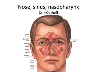
DEFINITION of PHARYNX • The pharynx is that part of the digestive tube which is placed behind the nasal cavities, mouth, and larynx. It is a wide musculomembranous tube, somewhat conical in form, with the base upward, and the apex downward, extending from the under surface of the skull to the level of the cricoid cartilage in front, and that of the sixth cervical vertebra behind .
NASOPHARYNX It lies behind the nose and above the level of the soft palate. It differs from the oral and laryngeal parts of the pharynx in that its cavity always remains patent. In front it communicates through the choanæ with the nasal cavities. On its lateral wall is the pharyngeal ostium of the auditory tube, somewhat triangular in shape, and bounded behind by a firm prominence, the torus or cushion,.Behind the ostium of the auditory tube is a deep recess, the pharyngeal recess (fossa of Rosenmüller). On the posterior wall is a prominence, best marked in childhood, produced by a mass of lymphoid tissue, the adenoids.
The adenoids • are a clump of lymphoid tissue similar to that of tonsils, but located higher up in the throat, behind the nose. Adenoids help the body fight infections by trapping and fighting micro organisms as they pass through the breathing passage.
Indications for adenoidectomy • Adenoidectomy is indicated if there is a chronic effusion in the middle ear in an adult, especially on one side only, which does not resolve relatively rapidly (3-6 weeks) with proper medical treatment. • Obstruction behind the nose causing snoring, airway obstruction, or poor sleep
Adenoidectomy Indicated when Enlarged adenoids are blocking the airway, which may be suspected if the child snores excessively has trouble breathing through the nose has episodes of not breathing during sleep The child has chronic ear infections that: interfere with child's education persist despite antibiotic treatment recur 5 or more times in a year recur 3 or more times a year during a 2-year period
Adenoidectomy • The adenoids normally shrink as the child reaches adolescence and adenoidectomy is rarely needed after reaching the teenage years. Adenoidectomy can done as an outpatient procedure in good set ups.. Complete recovery takes 1 to 2 weeks. While healing, the child may have a stuffy nose, nasal drainage, and a sore throat. Soft, cool foods and drinks may help relieve throat discomfort.
JUVENILE NASOPHARYNGEAL ANGIOFIBROMA
definition angiofibromas are highly vascular, non-encapsulated tumours affecting predominantly young males. These lesions are benign histologically but they may become life-threatening with excessive bleeding or intracranial extension.
JUVENILE NASOPHARYNGEAL ANGIOFIBROMA STUDY PERIOD – 1984 TO 2004 60 CASES ALL CASES EVALUATED ACCORDING TO A QUESTIONNAIRE FOLLOWUP – 18 MONTHS TO 4 YEARS PRE-OP TRACHEOSTOMY IN ALL PATIENTS
JUVENILE NASOPHARYNGEAL ANGIOFIBROMA ALL MALES AVERAGE AGE – 17 YEARS (RANGE 12-22 YRS) 60 CASES
JUVENILE NASOPHARYNGEAL ANGIOFIBROMA EPISTAXIS NASAL BLOCKAGE EAR COMPLAINTS FACIAL SWELLING COMMON PRESENTATION N = 60
JUVENILE NASOPHARYNGEAL ANGIOFIBROMA PRIMARY HAEMORRHAGE – 1 L (Ave) SECONDARY HAEMORRHAGE – 3 PATIENTS WOUND INFECTION – 3 PATIENTS CONDUCTIVE HEARING LOSS – 1 PATIENT HYPERTROPHIED SCAR – 3 PATIENT COMPLICATIONS
JUVENILE NASOPHARYNGEAL ANGIOFIBROMA FOLLOWUP 18 MONTHS TO 4 YEARS RECURRENCE 11 PATIENTS (18.3 %)
JUVENILE NASOPHARYNGEAL ANGIOFIBROMACONCLUSIONS SURGERY IS THE TREATMENT OF CHOICE MOST COMMON PRESENTATION IS EPISTAXIS BEST APPROACH IS TRANSPALATAL WITH LAT. RHINOTOMY FOLLOW UP CT SCAN AFTER 6 MONTHS