Download
1 / 21
210 likes | 367 Vues
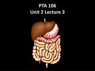
PTA 106 Unit 2 Lecture 3. Digestive Functions. Ingestion intake of food Digestion breakdown of molecules Absorption uptake nutrients into blood/lymph Defecation elimination of undigested material. Stages of Digestion. Mechanical digestion

E N D
Digestive Functions Ingestion intake of food Digestion breakdown of molecules Absorption uptake nutrients into blood/lymph Defecation elimination of undigested material
Stages of Digestion • Mechanical digestion • physical breakdown of food into smaller particles • teeth and churning action of stomach and intestines • Chemical digestion • series of hydrolysis reactions that break macromolecules into their monomers • enzymes from saliva, stomach, pancreas and intestines • results • polysaccharides into monosaccharides • proteins into amino acids • fats into glycerol and fatty acids
Subdivisions of Digestive System • Digestive tract (GI tract) • 30 foot long tube extending from mouth to anus • Accessory organs • teeth, tongue, liver, gallbladder, pancreas, salivary glands
Lesser and Greater Omentum • Lesser - attaches stomach to liver • Greater - covers small intestines like an apron
Stomach • Mechanically breaks up food, liquifies food and begins chemical digestion of protein and fat • resulting soupy mixture is called chyme • Does not absorb significant amount of nutrients • absorbs aspirin and some lipid-soluble drugs
Gross Anatomy of Stomach • Muscular sac (internal volume from 50ml to 4L) • J - shaped organ with lesser and greater curvatures • regional differences • cardiac region just inside cardiac orifice • fundus - domed portion superior to esophageal opening • body - main portion of organ • pyloric region - narrow inferior end • antrum and pyloric canal • Pylorus - opening to duodenum • thick ring of smooth muscle forms a sphincter
Liver, Gallbladder and Pancreas • All release important secretions into small intestine to continue digestion
Gross Anatomy of Liver • 3 lb. organ located inferior to the diaphragm • 4 lobes - right, left, quadrate and caudate • falciform ligament separates left and right • round ligament, remnant of umbilical vein • Gallbladder adheres to ventral surface between right and quadrate lobes
Ducts of Gallbladder, Liver, Pancreas • Bile passes from bile canaliculi between cells to bile ductules to right and left hepatic ducts • Right and left ducts join outside liver to form common hepatic duct • Cystic duct from gallbladder joins common hepatic duct to form bile duct • Duct of pancreas and bile duct combine to form hepatopancreatic ampulla emptying into duodenum at major duodenal papilla • sphincter of Oddi (hepatopancreatic sphincter) regulates release of bile and pancreatic juice
Ducts of Gallbladder, Liver, Pancreas • Bile passes from bile canaliculi between cells to bile ductules to right and left hepatic ducts • Right and left ducts join outside liver to form common hepatic duct • Cystic duct from gallbladder joins common hepatic duct to form bile duct • Duct of pancreas and bile duct combine to form hepatopancreatic ampulla emptying into duodenum at major duodenal papilla • sphincter of Oddi (hepatopancreatic sphincter) regulates release of bile and pancreatic juice
Small Intestine • Nearly all chemical digestion and nutrient absorption occurs in small intestine
Small Intestine • Duodenum curves around head of pancreas (10 in.) • retroperitoneal along with pancreas • receives stomach contents, pancreatic juice and bile • neutralizes stomach acids, emulsifies fats, pepsin inactivated by pH increase, pancreatic enzymes • Jejunum - next 8 ft. (in upper abdomen) • has large tall circular folds; walls are thick, muscular • most digestion and nutrient absorption occur here • Ileum - last 12 ft. (in lower abdomen) • has peyer’s patches – clusters of lymphatic nodules • ends at ileocecal junction with large intestine
Water Balance • Digestive tract receives about 9 L of water/day • .7 L in food, 1.6 L in drink, 6.7 L in secretions • 8 L is absorbed by small intestine and 0.8 L by large intestine • Water is absorbed by osmosis following the absorption of salts and organic nutrients • Diarrhea occurs when too little water is absorbed • feces pass through too quickly if irritated • feces contains high concentrations of a solute (lactose)
Gross Anatomy of Large Intestine • 5 feet long and 2.5 inches in diameter in cadaver • Begins as cecum and appendix in lower right corner • Ascending, transverse and descending colon frame the small intestine • Sigmoid colon is S-shaped portion leading down into pelvis • Rectum - straight portion ending at anal canal
Absorption and Motility • Transit time is 12 to 24 hours • reabsorbs water and electrolytes • Feces consist of water and solids (bacteria, mucus, undigested fiber, fat and sloughed epithelial cells • Haustral contractions occur every 30 minutes • distension of a haustrum stimulates it to contract • Mass movements occur 1 to 3 times a day • triggered by gastrocolic and duodenocolic reflexes • filling of the stomach and duodenum stimulates motility • moves residue for several centimeters with each contraction
Anatomy of Anal Canal • Anal canal is 3 cm total length • Anal columns are longitudinal ridges separated by mucus secreting anal sinuses • Hemorrhoids are permanently distended veins