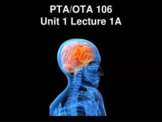

PTAOTA 106 Unit 1 Lecture 3
E N D
Presentation Transcript
The Basics • Arteries: Carry blood away from the heart toward tissues. They typically have thicker vessels walls to handle increased pressure. Contain internal and external elastic lamina that allow stretch and recoil (the reason you can feel a pulse clinically). • On models, arteries are colored red if they carry oxygenated blood and blue if they carry deoxygenated blood. • Veins: Carry blood back to the heart from tissues. They have thinner vessels walls designed to collapse and one way valves. They lack elastic lamina. Compression of veins by muscle contraction helps move blood. • On models, veins are blue if the carry deoxygenated blood and red if they carry oxygenated blood.
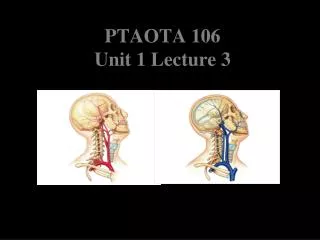
Major Arteries Superior Temporal Maxillary Facial Occipital Internal Carotid External Carotid Common carotid Vertebral Subclavian Brachiocephalic Trunk Major Veins Superior sagittal Sinus Transverse Sinus Sigmoid Sinus Temporal Occipital Facial Maxillary External Jugular Internal Jugular Vertebral Brachiocephalics Elements of the Cardiovascular System found in the head and neck region are the arteries and veins that supply the region
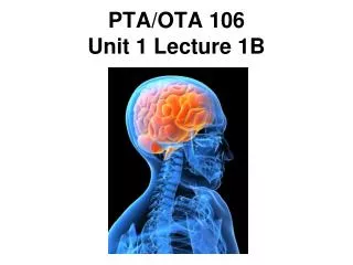
Clinical Concerns Occlusions:Obstruction of blood flow, result from thrombi, thrombophlebitis, or tumors. Cerebral contusions: brain trauma in which the pia is striped for the brain surface allowing blood to enter the subarachnoid space . Cerebral Lacerations:Damage that results in ruptured blood vessels allowing bleeding into the brain and subarachnoid space, causing intracranial pressure and cerebral compression. Ischemic Stroke: Impaired cerebral blood flow with development of neurological deficits. Most common causes are spontaneous cerebrovascular accidents such as embolism, thrombosis, hemorrhage, subarachnoid hemorrhage.
Clinical Concerns Hemorrhagic Stroke: Follows the rupture of an artery usually caused by an aneurysm. Transient Ischemic Attack (TIA): Neurological symptoms resulting from temporary ischemia. The symptoms of staggering, dizziness, light-headedness, fainting, and parasthesias last a few minutes, but can persist for up to an hour.