QUESTION
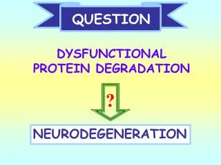
QUESTION. ?. NEURODEGENERATION. DYSFUNCTIONAL PROTEIN DEGRADATION. NEURODEGENERATION associated with Alzheimer’s Disease Parkinson’s Disease Huntington’s Disease Amyotrophic Lateral Sclerosis. Neurodegenerative Disorders. Disease. Parkinson’s Disease. Alzheimer’s Disease.

QUESTION
E N D
Presentation Transcript
QUESTION ? NEURODEGENERATION DYSFUNCTIONAL PROTEIN DEGRADATION
NEURODEGENERATION associated with Alzheimer’s Disease Parkinson’s Disease Huntington’s Disease Amyotrophic Lateral Sclerosis
Neurodegenerative Disorders Disease Parkinson’s Disease Alzheimer’sDisease accumulation of misfolded proteins Huntington’s Disease Amyotrophic lateral sclerosis Spinocerebellar Ataxia Cell Death
c d AD: tau f AD: ubiquitin PD: ubiquitin Ubiquitin-Protein Aggregates HUNTINGTON’S ALZHEIMER’S PARKINSON’S LOU GEHRIG’S
Protein Degradation • Turnover of protein is NOT constant • Half lives of proteins vary from minutes to infinity • “Normal” proteins – 100-200 hrs • Short-lived proteins • regulatory proteins • enzymes that catalyze committed steps • transcription factots • Long-lived proteins • Special cases (dentin, crystallins)
Protein Degradation • Proteins are not degraded at the same rate ENZYMEhalf-life Ornithine decarboxylase 11 minutes -Aminolevulinate synthetase 70 minutes Catalase 1.4 days Tyrosine aminotransferase 1.5 hours Tryptophan oxygenase 2 hours Glucokinase 1.2 days Lactic dehydrogenase 16 days HMG CoA reductase 3 hour
May depend on tissue distribution • Example: Lactic Acid Dehydrogenase • Tissue Half-life • Heart 1.6 days • Muscle 31 days • Liver 16 days • Protein degradation is a regulated process • Example: Acetyl CoA carboxylase • Nutritional state Half-life • Fed 48 hours • Fasted 18 hours Protein Degradation
Protein Degradation • Ubiquitin/Proteasome Pathway • 80-90% • Most intracellular proteins • Lysosomal processes • 10-20% • Extracellular proteins • Cell organelles • Some intracellular proteins
Two Sites for Protein Degradation • Proteasomes • Large (26S) multiprotein complex (28 subunits) • Degrades ubiquitinated proteins • Lysosomes • Basal degradation – non-selective • Degradation under starvation – selective for “KFERQ” proteins
The Ubiquitin/Proteasome PATHWAY
UBIQUITIN • Small peptide that is a “TAG” • 76 amino acids • C-terminal glycine - isopeptide bond with the e-amino group of lysine residues on the substrate • Attached as monoubiquitin or polyubiquitin chains • Three genes in humans: • Two are stress genes (B and C) • One, UbA as a fusion protein G K
Tetra-Ubiquitin Cook, W.J. et al. (1994) J. Mol. Biol. 236, 601-609
Ubiquitin/Proteasome Pathway Degradation by the 26S PROTEASOME Ubiquitination Ubiquitination
The Ubiquitin/Proteasome PathwayFour Main Steps: • UBIQUITINATION • RECOGNITION • DEGRADATION • DEUBIQUITINATION
UBIQUITIN CHAINS 6 11 27 29 33 MQIFVKTLTGKTITLEVESSDTIDNVKAKIQDKEGIPPDQQRLIFAGKQLEDGRTLADYNIQKESTLHLVLRLRGG 48 63
Functions of Ubiquitination • Mono-ubiquitination • Receptor internalization • Endocytosis – lysosome • Transcription regulation • Poly-ubiquitination • Targets proteins from Cytoplasm, Nuclear & ER for degradation by the PROTEASOME • DNA repair
Ubiquitination of proteins is a FOUR-step process • First, Ubiquitin is activated by forming a link to “enzyme 1” (E1). AMP • Then, ubiquitin is transferred to one of several types of “enzyme 2” (E2). • Then, “enzyme 3” (E3) catalizes the transfer of ubiquitin from E2 to a Lys e-amino group of the “condemned” protein. • Lastly, molecules of Ubiquitin are commonly conjugated to the protein to be degraded by E3s & E4s
UBIQUITIN ACTIVATION E1 UBIQUITIN ADENYLATE THIOL ESTER
N C UBC domain UBIQUITIN CONJUGATION E1-s-co-Ub + E2-SH -----> -----> E2-s-co-Ub+ E1 CLASS 1 – UBC domains only; require E3s for Ub; target substrates for degradation CLASS 2 – UBC domains & C-terminal extensions; UBC2 = RAD6 – DNA repair not degradation; no E3s CLASS 3 – UBC domains & N-terminal extensions; function not known
UBIQUITIN LIGATION E3 “recognins” =recognize a motif(DEGRON)on a protein substrate E2-s-co-Ub + Protein-NH2 -------> E2-SH + Protein-NH-CO-Ub (ubiquitin = polyubiquitin chains)
Three Major Classes of E3 1) HECT-domain E3s 3) multi-subunit cullin based E3s 2) RING finger-domain E3s
Ubiquitin Ligases (E3) • 1)HECT-domaincontaining a conservedCys • 2)RING finger-domain • Cys&Hisresidues are ligands to two Zn++ ions • stabilizes a molecularscaffold
Ubiquitin Ligases (E3) (cont.) • 3) Complex E3s: Multiple subunits • Ex: SCF-type E3, VBC-Cul2 E3 and other cullin based E3s, • Anaphase promoting complex (APC) • -they provide a Scaffold • for Ub transfer • -F-box – substrate • recognition
ELONGATION = E4 • U box = CHIP (+parkin) • Non-U box = p300 (p53) • E3-E4 complex = • C. elegans
RECOGNITION DEGRADATION SIGNALS substrates
N-end RULE N-degron - signal N-recognin - E3
PROTEASOME COMPONENTS 19-3 20S Proteasome 19S Particle ATP 26S Proteasome
Ubiquitinated proteins are degraded by the proteasome • Ubiquitinated proteins are degraded in the cytoplasm and nucleus by the proteasome. • Proteasomal protein degradation consumes ATP. • The proteasome degrades the proteins to ~8 amino-acid peptides. • Access of proteins into the proteasome is tightly regulated. • The peptides resulting from the proteasome activity diffuse out of the proteasome freely.
Hydrolysis peptide bonds after: hydrophobic a.a. =CHYMOTRYPSIN-LIKE - 5 acidic a.a. = (-) CASPASE-LIKE -1 basic a.a. = (+) TRYPSIN-LIKE -2
DEUBIQUITINATION De-ubiquitinating
Ubiquitin – like proteins “UBP” Small Ubiquitin-like Modifier
Digestive System of the Cell • Digests • ingested materials • obsolete cell components • Degrades macromolecules of all types • Proteins • Nucleic acids • Carbohydrates • Lipids • Heterogeneous
Lysosomal Enzymes • 50 different degradative enzymes • Acid hydrolases • Active at pH 5 (inside lysosome) • Inactive if released into cytosol (pH 7.2) • Acidic pH of lysosomes maintained by a proton pump in the lysosomal membrane • Requires ATP, thus mitochondria
Different pathways lead to the lysosome • 1) Phagocytosis • Cell “eating” of material • > 250nm • 2) Pinocytosis • Cell “drinking” • < 150nm • 3) Receptor Mediated • Endocytosis • -clathrin-coated pits • 4) Autophagy • “self eat” of old worn out organelles, • important in cell degradation duringapoptosis
Protein degradation in the lysosomes • Lysosomes degrade extracellular proteins that the cell incorporates by endocytosis. • Lysosomes can also degrade intracellular proteins that are enclosed in other membrane-limited organellas. • In well-nourished cells, lysosomal protein degradation is non-selective(non-regulated). • In starved cells, lysosomes degrade preferentially proteins containing a KFERQ“signal” peptide. • The regression of the uterus after childbirth is mediated largely by lysosomal protein degradation
AUTOPHAGY • - Macroautophagy – inducible (mTOR) • (autophagy) • - Microautophagy - constitutive • - Chaperone-mediated autophagy • (CMA) – KFERQ motif