Download
1 / 20
200 likes | 219 Vues
Learn about the general characteristics of mycobacteria, their acid-fast nature, and the different types of tuberculosis. Explore the pathogenicity, diagnosis, and treatment of tuberculosis.

E N D
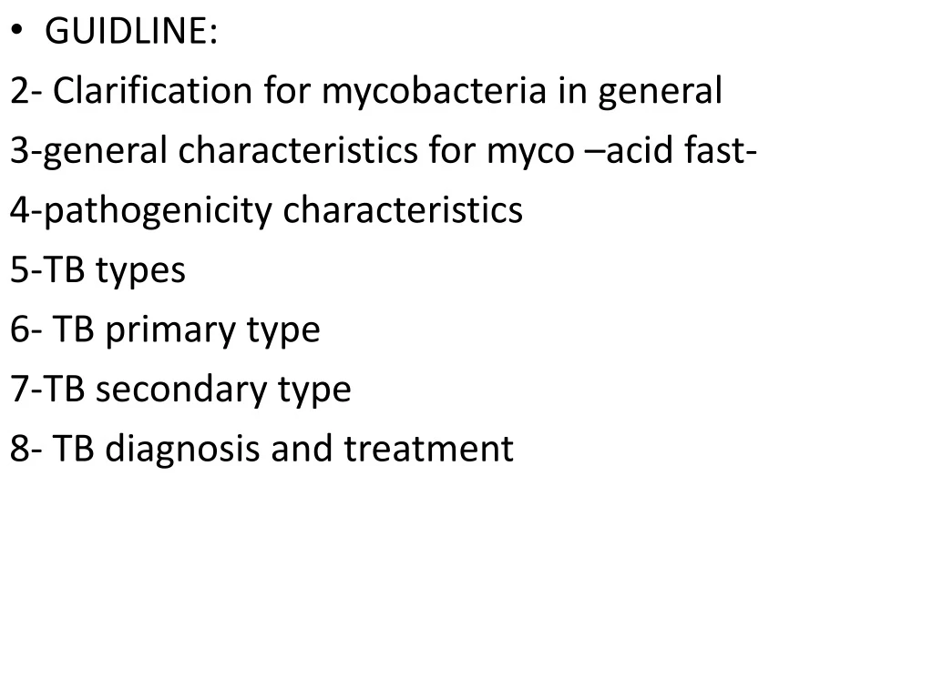
GUIDLINE: 2- Clarification for mycobacteria in general 3-general characteristics for myco –acid fast- 4-pathogenicity characteristics 5-TB types 6- TB primary type 7-TB secondary type 8- TB diagnosis and treatment
Mycobacteria (Acid-Fast) • Mycobacteria is divided into 3 types: • M.tuberculosis • M.Leprae –we didn’t take- • NontuberculosisMycobacteria –we didn’t take- They are rods with lipid-laden cell walls, and that’s what makes them acid fast, how? When we have a smear of sputum for example, and we cover it with red stain carbofusion -stain with high affinity for mycobacterial cell walls component, it’s found in Ziehl-Neelsen stain -and heated for better penetration, then poured with acid alcohol -95% ethanol and 3% HCL- and then counterstained with methylene blue, the cell wall lipids will not dissolve when alcohol is applied and thus red stain won’t be washed off. So acid-fast organisms resist decolorization and hold fast to their red stain.
Mycobacteria Group Acid-Fast Bacilli: • are obligate aerobic which makes snese because they mostly infect the lungs where O2 is abundant • Their cell callcontains protein-polysaccharides with high Phospholipids (mycolic acid –large fatty acid-, waxes) and they act like virulence factors (it is loaded with lipids and that’s what makes them acid-fast ). • When they attack tissues they cause Necrosis, in case of tuberculosis as we took in pathology they cause caseous necrosis and forms granuloma where they can remain viable. • Resistant to Dryness, low Acidity, Alcohol and detergents (like carbafusion mentioned before) • Susceptible to UV-light and heat. • They’re Common in Human and infected ones are Asymptomatic persons -like in case of primary asymptomatic tuberculosis which is the common type but the primary symptomatic is less common and happen mostly in children and elderly or immunocompromised patients because they have a weak immune system or when reactivation happens- , domestic Animal and Birds, Environment • They kill 3-5 Million yearly.
Human/animals Pathogens: • Slow growth in vitro, the culture needs (2-6 weeks) • Nonpathogenic species: lives in genital tract or skin ( like M. smegmatis which has rapid growth 3-7 days and lives in normal genital secretions). • Pathogenic species : Mostly M. tuberculosis and few percentage M. boviswhich causes tuberculosis in Animals –cattle- , and if transmitted through Dairy products to humans can cause Intestinal tuberculosis. Atypical Mycobacteria (nontuberculosismycobacteria) they are divided into pigmented and non-pigmented, common in environment and Rarely cause lung Tuberculosis.
Pulmonary Tuberculosis: • Exudative, primary type • Active productive, reactivation, secondary type
Pulmonary Tuberculosis/ Exudative type –primary-: • Slow intracellular growth in lung tissue • Incubation time 1-12 months transmitted via droplet infection (cough,laugh,..) • Primarily causes mild Lung lesion infects Mostly Children (90%) • Causes Asymptomatic infection discussed earlier and Rarely causes active lesions which are called (cavitary lesions with air-fluid levels) caused by progressive tuberculosis leading to severe cases where lung necrosis is developed and cavities and holes are formed and they’ll be filled with fluid which is seen in CT scan and chest radiography. • Recovery: even with no treatment most cases will control infection and have healing by encapsulation and forming granuloma which decrease the number but they’re still viable. • Positive skin tuberculin test: it’s anintradermalinjecetion of antigenic protein particles from killed M.tuberculosis, will reveal if the person is infected or not, because many of infected individuals will not manifest a clinical infection for years. When a positive PPD test occurs, you can treat and eradicate the disease before it significantly damages the lungs or other organs. And we use it when we have suspects in person with a low-grade fever and cough or a person who has been in contact with infected ones. We inject it intradermally and within 1-2 days, skin will be red, raised and hard (positive)
7. Hypersensivity Immunity “ Asymptomatic infection is not necessary result in Disease.” • Active-Productive type: Adult infection: Most adult cases of tuberculosis occur after the bacteria have been dormant for some time. • Happens by Reactivation of old tuberculosis lesions • may present in any Body site, The infection can occur in any of the organ systems seeded during the primary infection. It is presumed that a temporary weakening of the immune system may precipitate reactivation. Like in Intestinal tract, Kidney and bones. • Meningitis common in children: TB causes subacute meningitis and forms granulomas in the brain. • Lung lesion: patient will usually persent with a chronic lowgrade feverCough, Bloody sputum, night sweats and weight loss. • Detection by X-ray and shows positive tuberculin test, Larger reaction.
Lab Diagnosis: • Direct AFS(acid fast stain) : Ziehl-Neelsen stain • Culture in Lowenstein -Jensen Medium from Sputum, urine, Pleural fluid, CSF, Biopsy.we have to incubate for at least 6 weeks. However we may recognize the first colonies after 2 weeks. From these colonies we have to prepare an Acid Fast Stain and then do biochemical tests to confirm a case of TB. • PPD • X-rays • Treatment: Combination of anti-tuberculosis drugs for 6-24 months. (Rifampin, streptomyocin,Isoniazid,..) • Prevention by BCG vaccine( Bacilli Calemtte- Guerin) for Children.
Chlamydia group • Chlamydia Cell is Small and it has a Gram-ve wall but unlike other G-ves it has few amount of liposaccharides and no murmaric acid. • It’s obligate intracellular means it lives inside the cells and take ATP from host cells. • Dimorphic growth: has two forms 1)Infectious stage(Elementary bodies/ Infectious) which are responsible for attaching to the host mucosa cell and promoting its entry and then inhibit phagosome-lysosome fusion and develop into 2) (Inclusion bodies/Reticulate bodies) and replication occurs by binary fission. • There are 3 species only two are required: • ChlamidyaTrachomatis • ChlamidyaPneumoniae
Firstly, Chlamydia trachomatis: infects eyes and genital organs Genital tract: A common cause of STD worldwide causes Nonspecific urethritis –urethritisisi caused by nisseria also and called gonococcalurethritis but the one caused by chalmydia is nongonococcalurethritis that’s why it’s called NGU or nonspecific only for differentiation- and it’s associated with pus. It can also cause Prostatitis, Vagnitis and Cervicitiswhich are associated with discharges and the inflamed areas are swollen and red, it might lead to infertility Eyes: Newborns with chlamydial conjunctivitis: Pregnant mothers during pregnancy especially in the delivery can transmit the disease to her fetus, infecting its Eyes causing an inflammatory reaction in the conjunctiva and later in the cornea, producing trachoma; trachoma means developing damage in the conjuctiva causing blindness; and this is associated with blindness so it can be so severe. All newborns in the US are given erythromyocin eye droplet prophylatically. infected patients shows mild to severe eyes redness, swollen eyelids and discharge from the eyes which can be thin and watery or thick and yellow.. Trachoma leads to Blindness if not treated.
Secondly, Chlamydia pneumoniae: Attached to Tracheal Epithelial cells and cause acute bronchitis and Atypical pneumonia which is Mild-severe pulmonary infection associated with mild-sever dry cough, abdominal paing and some GI symptoms and it may recover without the need of antibiotic –not dangerous or fatal- and it’s Common in children in All ages • Diagnosis & treatment : it’s diagnosed by Clinical features & serological test. • After 4-8 weeks of chlamidya incubation is develops antibodies against it and then could be discovered • It’s cultured only in McCoy tissue culture –livinng culture- because it can’t live in nonliving cultures because of the need of ATP from the host cells. • PCR test are also used and immunoflourescence techniques. • Treated by Antibiotics and there’s No Vaccine
Mycoplasma group • The smallest Bacteria and what is unique about it that it Lacks Cell Wall but has a Lipid bi-layer Membrane paded with cholestrol coating it –so penicillin cannot attack it because its target is peptidoglycans in cell wall which not found-. • Aerobic and lives in Respiratory/Urinary Mucosa. • Found in Human, Animals and Birds. • It has 2 species : • M. pneumoniae: Human pathogens. causes mildPharyngitis, Bronchitis, Pneumonia, and associated with Dry cough and Fever –similar to atypical pneumonia-. Most Common in old children & Young adults and it’s Less Elderly, Common infection in Fall-Winter. • M.hominis/M.genitalium:Part oforal-genital floracause Nonspecific Urethritis, Vaginitis, Cervicitis. • Diagnosis & treatment:Sputum, Urine Culture, Cold-Agglutination Test, ELSA Specific antibodies, PCR. TREATMENT : Antibiotics and No Vaccine.
Legionella pneumonphila • It causes Legionnaires’ disease –severe pneumonia- which was found in 1976 USA • It’s Thin G-ve Coccobacilli-Filments. • Facultative Anaerobes and Survive at 0-80 C. • Lives in Cold/Hot Water, Air Condition, Wet Soil, Aerosols بخاخاتFine sprays. • Droplet infection. • Infect Respiratory Mucosa. • It’s a facultative Intracellular parasite that settle in the lower RT and is globbed up by Monocyte-Macrophage This means that once it’s phagocytosed it inhibits the phagosom-lysosome fusion, surviving and replicating intracellularly • Extracellular growth,Not contagious disease.
Legionella Pneumophila • Clinical Features:High Fever, dry Cough, vomiting, stomach discomfort and Diarrhea. Other common symptoms include headaches, muscle aches, chest pain, and shortness of breath, Pneumonia, Renal Failure, and if it was severe it might lead to Death. • Mostly common in elderly, Immumodeficient patients like AIDS pateints and heavy Smoking Persons. • Diagnosis & treatment:Special Culture Media, Blood-sputum culture. Detection by Specific antibodies, PCR. Treatment: Antibiotics.. No Vaccine. Legionellapneumophila is the most common cause of community acquired pneumonia.
Spirochetes Group-1 • They’re Gram-ve and haveSpiral forms and Long –very selinder and tightly coiled- and Have long helically coiled cells (5-20um). • Common Human, Animals, Arthropodes. • Nonpathogenic /Pathogenic. • 2 species: • Treponema species: Nonpathogenic, and lives in Oral cavity. • Treponemapalldium: causes Syphilis which is a Veneral Disease(STD) transmitted through Sexual Contact. entering the host via breaches in squamous or columnar epithelium. • It gains access to host's blood and lymph systems through tissue and mucus membranes. • Incub. 2-week-Few Months: it changes from Acute to Chronic Infection with time (because it has stages)
Associated with Mucosa/Skin Lesions-Chancre on Genitalia, Anal area and Mouth. It’s a Systemic Disease and can Affect Any Body Organ because of its ability to reach blood and lymhpand causes Meningitis, Hepatitis, Nephritis, Granulomatous lesions. • Congenital Syphilis: happens with infected pregnant women, the Treponemapallidum crosses placenta and infect the fetus. • Diagnosis: spirochetes cannot be cultured in ordinary media and even it’s G-ve but it’s too small to be seen with light microscope so we use special ways like Direct Dark-field Microscopy, immunoflurecense and silver stain. • Serological Test like • VDRL (Venereal Disease Research Lab). • Fluorescent Trep. Antibody-Test (FTA). • No Culture and treated with Antibiotics
Spirochetes Group-2/ Borrelia 1- Borrelia Burgdorferi: causes Lyme Disease and Common in USA. • Carried by Biting Insects (Ticks), Wild Animals, Rodents and Birds. • Incubation : Few Weeks- Months. • The primary stage is featured by Single/Multiple Skin Erythematic Lesions and then in the upcoming stages after years it turns into a Systemic Disease by invading body systems and organs, and in stage 2 and 3 causing Arthritis, CNS diseases –encephalopathy and meningitis and nevre palsies- and Cardiac Abnormalities. 2- Borrelia Recurrentis is found Worldwide. They cause an Epidemic/Endemic Relapsing Fever by Biting Insects (Human Lice/ Animal Ticks) which may lead after reachin blood to Septicemia and associated with Low-High Fever, Chills, Severe Headache and symptoms are resolved in 3-6 days then developing similar features for another 3-6 dadys and relapses will continue to occur with shorter and milder inervals, Common Relapses. 3- Leptospira which causes Liptospiral diseases: Zoonosis –infect humans and animals- ,and cause mild-severe fatal systemic disease which is Weils’s disease(infection with jaundice), it involves renal failure, hepatits , mental status change and hemorrhage in many organs associated with high Fever, vasculitis , Bleeding. • Diagnosis: Serological Tests and Special fluid culture methods. Especially for CSF