Download
1 / 59
610 likes | 841 Vues
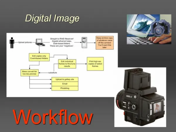
The Nature of the Digital Image. Ehsan Samei and Robert S. Saunders Digital Imaging Research Division, Department of Radiology, Physics, and Biomedical Engineering. Sept. 18, 2007. Introduction. Why Digital Imaging?

E N D
The Nature of the Digital Image Ehsan Samei and Robert S. Saunders Digital Imaging Research Division, Department of Radiology, Physics, and Biomedical Engineering Sept. 18, 2007
Introduction • Why Digital Imaging? • To facilitate more convenient distribution, display, and storage of medical images electronically, • To enable manipulation of image appearance by post-processing Visible light Computed radiography (CR) Screen-film system Charge-coupled devices (CCDs) Image intensifier (II) tube Complementary Metal-Oxide Semiconductor (CMOS) Flat panel detectors • Several important image quality concepts for digital X-ray imaging
Digital versus Analog Photo-conductor Scintillator optical glue film Pixel Picture element Discretization
Properties of material used for room temperature semiconductor detectors Material Cd0.9Zn0.1Te CdTe Si GaAs HgI2 PbI2 a-Si a-Se Atomic numbers 48,30,52 48,52 14 31,33 80,53 82,53 14 34 Average atomic number 49.1 50 14 32 62 62.7 14 34 Density r (g/cm3) 5.78 5.85 2.33 5.32 6.6 6.2 2.3 4.3 1.572 1.5 1.12 1.43 2.13 2.32 1.8 2.2 Bandgap Eg(eV) 10.9 11 11.7 12.8 8.8 11.7 6.6 Dielectric constant Pair creation energy Epair (eV) 4.64 4.43 3.62 4.2 4.2 4.9 4 47 Resistivity r (W cm) 3×10¹⁰ 109 < 104 107 1013 1012 1012 1012 Electron lifetime-mobility product mete (cm2/V) 5×10⁻³ 3×10⁻³ > 1 8×10⁻⁵ 10-4 8×10⁻⁶ 6×10⁻⁸ 5×10⁻⁹ Hole lifetime-mobility product mhth (cm2/V) 5×10⁻⁵ 2×10⁻⁴ 1 4×10⁻⁶ 4×10⁻⁵ 2×10⁻⁸ 1×10⁻⁷
Material Advantage Disadvantage Cd0.9Zn0.1Te chemically stable, good charge transport limitation in large area application (2 inch), high costs CdTe excellent charge transport poor radiation hardness Si relatively large area (6 inch) high leakage current GaAs chemical and mechanical stability (weak at moisture and brittle) low leakage current, low growth temperature HgI2 limited sensitivity due to charge carrier Schubweg limitation low leakage current, low growth temperature PbI2 excellent charge transport, large area poor radiation hardness a-Si a-Se low leakage current, low growth temperature high pair creation energy (47 eV), recrystallization at room temp.
Intensity discretization process : maximum contrast resolution
Basic Descriptor of the Digital Image(Image Size) • Desired imaging application ( 12 to 43 cm) Mammography applications: 18 x 23, 25 x 29 cm2, General radiography and chest applications : 43 x 43 cm2 Hologic Selenia Sterling 19” x 17” ( 43 x 35 cm 2 ) ( 3k x 2.5k ) Trixell Co. ( in France ) Thomson, Philips, Siemens 17”x 17” ( 3k x3k )
Basic Descriptor of the Digital Image(Latitude, Dynamic range) • : the range over which the detector is able to provide clinically useful images. E max : the exposure at which the detector saturate E min : the exposure at which the image pixel value just exceeds the background instrumentation noise k : how much the signal amplitude must exceed noise for reliable detection.
Basic Descriptor of the Digital Image(Latitude, Dynamic range) E max E max E min object detector E max E min E min
Basic Descriptor of the Digital Image(Latitude, Dynamic range) Exposure range (E max/E min) Mammographic imaging Radiography/fluoroscopic imaging
Basic Descriptor of the Digital Image(Spatial Resolution) • ability of the imaging system to present details in the objects being imaged. ( small or subtle) • MTF (modulation transfer function) • The ability of an imaging system to transfer a single input spatial frequency component, or sinusoid, into an output sinusoid of the same frequency in terms of relative signal amplitude of the output sinusoid. • Digital detector’s limiting frequency p: pixel size
Basic Descriptor of the Digital Image(Spatial Resolution) input output
Basic Descriptor of the Digital Image(Spatial Resolution) Ghosting artifacts - Control of pixel size - Limits of limiting frequency
Basic Descriptor of the Digital Image(Spatial Resolution) • Pre-sampled MTF • the detector spatial frequency response before the sampling stage • The edge and slit technique • The edge technique is international standard (IEC2003)
화면 캡처: 2007-09-18, 오후 4:56 Basic Descriptor of the Digital Image(Spatial Resolution) Edge method Slit method Tantalum phantom, 1.5 mm thickness 10 um slit width Lead foil, 250 um thickness
화면 캡처: 2007-09-18, 오후 4:56 Basic Descriptor of the Digital Image(Spatial Resolution) Edge method Edge spread function Differentiation and then Fourier transform Line spread function MTF
Edge Image Ideal case Practical case
화면 캡처: 2007-09-18, 오후 4:56 Basic Descriptor of the Digital Image(Spatial Resolution) Edge spread function Line spread function MTF
화면 캡처: 2007-09-18, 오후 4:56 Basic Descriptor of the Digital Image(Spatial Resolution) Presampled MTF
화면 캡처: 2007-09-18, 오후 4:56 Basic Descriptor of the Digital Image(Spatial Resolution) Slit method
화면 캡처: 2007-09-18, 오후 4:56 Basic Descriptor of the Digital Image(Spatial Resolution) Fourier Transform Line spread function MTF
Basic Descriptor of the Digital Image(Noise) • any image feature that impairs a specified diagnostic task • anatomical noise, • radiographic noise : image fluctuations added during image acquisition • Quantum noise : Poisson statistics of X-ray detection • Instrumentation noise : detector electronics, detector response non- uniformity. • NPS (noise power spectrum) • The spatial frequency content of noise • Fourier transform of the autocorrelation function (ACF) • ACF : characterizing the correlation of noise fluctuations in a uniform image
Basic Descriptor of the Digital Image(Noise) Example of NPS calculation pattern profile of autocorrelation autocorrelation
Basic Descriptor of the Digital Image(Noise) Image carried by X-ray Quanta Recoded Image Signal Output Signal Input Detector Noise Input Noise Output
Basic Descriptor of the Digital Image(Efficiency) The quality of a digital image : signal to noise ratio (SNR) G : system gain NEQ : noise equivalent quanta, exposure dependent DQE : How efficiently an imaging system utilizes an input X-ray flux DQE : detective quantum efficiency, how the imaging system transmits an input SNR to the output image SNR Q : the square of the ideal signal to noise ratio
Basic Descriptor of the Digital Image(Noise) Image carried by X-ray Quanta Recoded Image Signal Output Signal Input Detector Noise Input Noise Output
Basic Descriptor of the Digital Image(Scatter Sensitivity) Scatter : the process in X-ray imaging in which the X-ray photons are randomly deflected from their incident direction Uniform scattering : lowers image contrast Scatter varies across an image and contributes to total image noise (due to the difference of density in anatomy)
Basic Descriptor of the Digital Image(Scatter Sensitivity) Scattered fraction as high as 90 % • The main approach to reducing the scattered radiation • 1) anti-scatter grid : common method, scatter fraction 68 % • 2) the air gap: no impact on primary radiation, spreading • of the primary photons over a large area • 3) scanning slot imaging: a narrow detector scans across • the patient eDQE (effective detective quantum efficieny) t : the primary transmission through the grid SF : the scatter fraction
Basic Descriptor of the Digital Image(Speed) Speed : the radiographic exposure necessary to obtain an adequate quality image Analog screen-film radiography : the exposure necessary to achieve a certain optical density and const Ks : air kerma in mGy to achieve an optical density of one K0 : 1 mGy = 0.57 mR Ex) speed 400
Basic Descriptor of the Digital Image(Speed) Digital radiography : image gray-value and contrast is no longer limited by the applied exposure. the exposure required to obtain certain a certain SNR in an acquired image S0 : the speed eDQE0(0) : zero-frequency effective DQE of a typical reference screen –film system Ex) 120 kVp, 12:1 greed with 50% transmission, a scatter fraction of 0.4, a reference 400 speed film with 25% DQE (0)
Basic Descriptor of the Digital Image(Lag) Lag : signal from a previous image is still present in a subsequent image In complete erasure in CR, finite decay times of the detector phosphor Incomplete charge collection from photodiode or photoconductor Trapped charge in photoconductor • Introduction of scrub or dark frame into the image acquisition process 2) Subtracting a percentage of the prior image in the image acquisition sequence
Basic Descriptor of the Digital Image(Spatial artifacts) Defective or dead pixel elements Inactive area that results from tiling smaller detector together into a large-area detector
An Overview of Digital Imaging System for Radiology and Fluoroscopy Michael V. Yester, Ph.D University of Alabama at Birmingham Department of Radiology Birmingham, Alabama
Important Imaging Considerations Receptor ( same with conventional films) - Spatial resolution, contrast, and dose efficiency Indirect – emission of light and subsequent capture of light to form the signal Direct – electron hole pair generation, charge collection Digitization
Important Imaging Considerations Film /Screen System Digital system Spatial resolution Contrast Dose efficiency A series of trade-offs
Receptor Properties 1) Fill factor – the percent of the active area with in a pixel (indirect DR system) Photosensitive area Active amplifier
Receptor Properties (direct DR system)
Receptor Properties (direct DR system) electrode X-ray receptor (a-Se or CdZnTe) bumping Amplifier and TFT(thin film transistor)
Receptor Properties 2) The fraction of absorbed radiation - Photoelectric interaction strongly dependent on the atomic number (Z) t I0 I • I0: Incident intensity , • I : intensity after penetration • m: linear attenuation coefficient • r: density of material • ma: atomic weight material, • Z : atomic number of material • : X-ray absorption cross section, • E : X-ray energy
Receptor Properties X-Ray Mass Attenuation Coefficients ( m/r) : X-Ray Mass Energy-Absorption Coefficients (men/r) : Calculation of Photon Mass Energy-Transfer and Mass Energy-Absorption Coefficients Stephen M. Seltzer Radiation Research, Vol. 136, No. 2. (Nov., 1993), pp. 147-170. http://physics.nist.gov/PhysRefData/XrayMassCoef/cover.html
Interaction with X-ray the incident x ray interacts with an electron in the medium interaction with all the electrons of the atom Figure 1.14: In Rayleigh scattering, the incident x ray interacts with the electric field of an orbiting electron and is scattered as a result. The energy of the scattered x ray (E') = the incident x ray (Eo). No ionization occurs in Rayleigh scattering. Rayleigh scattering is most likely for low-energy x rays and for high-Z absorbers. Figure 1.13: In the photoelectric effect, an x ray with energy Eo is absorbed by an atomic electron, which is ejected from the atom causing ionization. The photoelectron will have kinetic energy equal to Eo-EBE,
Photoelectric Absorption & Characteristic Radiation • Coherent Scattering ( Non-Ionizing Radiation )
Interaction with X-ray • The interaction of an incident x ray with the electric field of the nucleus. - Interaction with the free electron of atom - Interact near a nucleus in an attenuation media Figure 1.19: Pair production can occur when an incident x ray (with Eo > 1.02 MeV) interacts with the electric field of an atom. A negatron (e-)-positron (e+) ion pair is formed in the interaction. Pair production does not occur at diagnostic x-ray energies. Figure.1.17: an incident x ray with energy Eo interacts with an outer-shell electron. The electron is ejected from the atom, causing ionization. A scattered x-ray photon with energy E' emerges at an angle q relative to the incident photon's trajectory.
Interaction with X-ray Figure 1.21: The positrons produced in pair and triplet production will lose their kinetic energy by interaction with the medium, and then rapidly interact with any available negative electron (negatron) and annihilate, producing two 511-keV photons being emitted in opposite directions. The photons produced are called annihilation radiation. Figure 1.20: Triplet production occurs when an incident x ray (Eo> 2.04 MeV) interacts with the electric field surrounding an orbital electron. The orbital electron is ejected from the parent atom, along with a negatron/positron pair, resulting in three particles being emitted.
Mass attenuation coefficient Figure 1.24: The mass attenuation coefficient for iodine is illustrated as a function of x-ray energy. The K edge of iodine (at 33 keV) and the L edge (at 5.2 keV). Figure 1.25: The mass attenuation coefficient of lead is shown as a function of x-ray energy. The Kedge (88 keV), L edges (around 16 keV), and M edge (-3 keV). 5.2keV 33keV t=s, 200 t=s, 500
Mass attenuation coefficient Figure 1.26: The energy of various absorption edges is illustrated as a function of the atomic number of the element. For elements below Z = 10 (i.e., tissue), the K edges are below 1 keV. The placement of the K edge of an x-ray detector has a relatively important role to play in the detection properties of the system.