Download
1 / 1
10 likes | 134 Vues
Monitoring Chromosome Dynamics in a Living Bacterial Cell. Monica Olvera, Northwestern University, DMR 0520513.

E N D
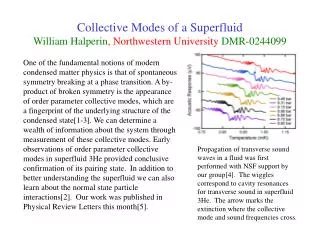
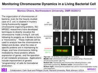
Monitoring Chromosome Dynamics in a Living Bacterial Cell Monica Olvera, Northwestern University, DMR 0520513 The organization of chromosomes of bacteria, even for the heavily studied case of E. coli, is cloaked in mystery. Using fluorescently-tagged chromosome-organizing proteins, NU-MRSEC researchers have developed techniques to directly visualize the chromosome inside a living E. coli cell, following its progeny as it divides to form a “microcolony”. These methods will be used to analyze how the chromosome is folded and divides, what the roles of specific proteins are in maintaining its shape, and how chromosome folding mediated by those proteins affects expression of genes, and the operation of cell metabolic processes. Applications include improvement of genetic “programming” of cells for biomaterial production. DIC (white light) chromosome (gfp-Fis) 1µm 0 min 10 20 25 30 membrane chromosome (gfp-Fis) in cell isolated Top row: cell dividing, early stage of microcolony. Middle row: chromosome inside cell visualized using a fluorescent chromosome protein (gfp-Fis). Bottom left: membrane and chromosome following division, note asymmetric positioning. Bottom center: chromosome in cell; Bottom right: chromosome isolated from cell for micromanipulation study; note expansion. Collaborators: Calin Guet and Philippe Cluzel, Harvard University; Reid Johnson, UCLA.