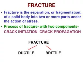
Fracture Classification
Fracture Classification. Lisa K. Cannada MD Revised: May 2011 Created March 2004; Revised January 2006 & Oct 2008. History of Fracture Classification. 18 th & 19 th century History based on clinical appearance of limb alone. Colles Fracture Dinner Fork Deformity. 20 th Century.

Fracture Classification
E N D
Presentation Transcript
Fracture Classification Lisa K. Cannada MDRevised: May 2011Created March 2004; Revised January 2006 & Oct 2008
History of Fracture Classification • 18th & 19th century • History based on clinical appearance of limb alone Colles Fracture Dinner Fork Deformity
20th Century • Classification based on radiographs of fractures • Many developed • Problems • Radiographic quality • Injury severity
What about CT scans? • CT scanning can assist with fracture classification • Example: Sanders classification of calcaneal fractures
The Soft Tissues Fracture appears non complex on radiographs The real injury
Patient Variables • Age • Gender • Diabetes • Infection • Smoking • Medications • Underlying physiology
Injury Variables • Severity • Energy of Injury • Morphology of the fracture • Bone loss • Blood supply • Location • Other injuries
Why Classify? • As a treatment guide • To assist with prognosis • To speak a common language with other surgeons
As a Treatment Guide • If the same bone is broken, the surgeon can use a standard treatment • PROBLEM: fracture personality and variation with equipment and experience
To Assist with Prognosis • You can tell the patient what to expect with the results • PROBLEM: Does not consider the soft tissues or other compounding factors
To Speak A Common Language • This will allow results to be compared • PROBLEM: Poor interobserver reliability with existing fracture classifications
Interobserver Reliability Different physicians agree on the classification of a fracture for a particular patient
Intraobserver Reliability For a given fracture, each physician should produce the same classification
Descriptive Classification Systems • Examples • Garden: femoral neck • Schatzker: Tibial plateau • Neer: Proximal Humerus • Lauge-Hansen: Ankle
Literature • 94 patients with ankle fractures • 4 observers • Classify according to Lauge Hansen and Weber • Evaluated the precision (observer’s agreement with each other) Thomsen et al, JBJS-Br, 1991
Literature • Acceptable reliabilty with both systems • Poor precision of staging, especialy PA injuries • Recommend: classification systems should have reliability analysis before used Thomsen et al, JBJS-Br, 1991
Literature • Classified identical 22/100 • Disagreement b/t displaced and non-displaced in 45 • Conclude poor ability to stage with this system • 100 femoral neck fractures • 8 observers • Garden’s classification Frandsen, JBJS-B, 1988
OTA Classification • There has been a need for an organized, systematic fracture classification • Goal: A comprehensive classification adaptable to the entire skeletal system! • Answer: OTA Comprehensive Classification of Long Bone Fractures
With a Universal Classification… You go from x-ray…. To… Treatment Implant options Results
To Classify a Fracture • Which bone? • Where in the bone is the fracture? • Which type? • Which group? • Which subgroup?
Using the OTA Classification • Where in the bone? • Which bone?
Proximal & Distal Segment Fractures • Type A • Extra-articular • Type B • Partial articular • Type C • Complete disruption of the articular surface from the diaphysis
Diaphyseal Fractures • Type A • Simple fractures with two fragments • Type B • Wedge fractures • After reduced, length and alignment restored • Type C • Complex fractures with no contact between main fragments
Grouping-Type A • Spiral • Oblique • Transverse
Grouping-Type B • Spiral wedge • Bending wedge • Fragmented wedge
Grouping-Type C • Spiral multifragmentary wedge • Segmental • Irregular
Subgrouping • Differs from bone to bone • Depends on key features for any given bone and its classification • The purpose is to increase the precision of the classification
OTA Classification • It is an evolving system • Open for change when appropriate • Allows consistency in research • Builds a description of the fracture in an organized, easy to use manner
Classification of Soft Tissue Injury Associated with Fractures
Closed Fractures • Fracture is not exposed to the environment • All fractures have some degree of soft tissue injury • Commonly classified according to the Tscherne classification • Don’t underestimate the soft tissue injury as this affects treatment and outcome!
Closed Fracture Considerations • The energy of the injury • Degree of contamination • Patient factors • Additional injuries
Tscherne Classification • Grade 0 • Minimal soft tissue injury • Indirect injury • Grade 1 • Injury from within • Superficial contusions or abrasions
Tscherne Classification • Grade 2 • Direct injury • More extensive soft tissue injury with muscle contusion, skin abrasions • More severe bone injury (usually)
Tscherne Classification • Grade 3 • Severe injury to soft tisues • -degloving with destruction of subcutaneous tissue and muscle • Can include a compartment syndrome, vascular injury Closed tibia fracture Note periosteal stripping Compartment syndrome
Literature • Prospective study • Tibial shaft fractures treated by intramedullary nail • Open and closed • 100 patients Gaston, JBJS-B, 1999
Literature What predicts outcome? Classifications used: • AO • Gustilo • Tscherne • Winquist-Hansen (comminution) All x-rays reviewed by single physician Evaluated outcomes Union Additional surgery Infection Tscherne classification more predictive of outcome than others Gaston, JBJS-B, 1999
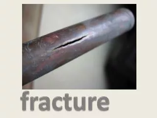
Open Fractures • A break in the skin and underlying soft tissue leading into or communicating with the fracture and its hematoma
Open Fractures • Commonly described by the Gustilo system • Model is tibia fractures • Routinely applied to all types of open fractures • Gustilo emphasis on size of skin injury
Open Fractures • Gustilo classification used for prognosis • Fracture healing, infection and amputation rate correlate with the degree of soft tissue injury by Gustilo • Fractures should be classified in the operating room at the time of initial debridement • Evaluate periosteal stripping • Consider soft tissue injury
Type I Open Fractures • Inside-out injury • Clean wound • Minimal soft tissue damage • No significant periosteal stripping
Type II Open Fractures • Moderate soft tissue damage • Outside-in mechanism • Higher energy injury • Some necrotic muscle, some periosteal stripping
Type IIIA Open Fractures • High energy • Outside-in injury • Extensive muscle devitalization • Bone coverage with existing soft tissue not problematic Note Zone of Injury
Type IIIB Open Fractures • High energy • Outside in injury • Extensive muscle devitalization • Requires a local flap or free flap for bone coverage and soft tissue closure • Periosteal stripping
Type IIIC Open Fractures • High energy • Increased risk of amputation and infection • Major vascular injury requiring repair
Literature on Open Fracture Classification • 245 surgeons • 12 cases of open tibia fractures • Videos used • Various levels of training (residents to trauma attendings) Brumback et al, JBJS-A, 1994
Literature on Open Fracture Classification • Interobserver agreement poor • Range 42-94% for each fracture • Least experienced-59% agreement • Orthopaedic Trauma Fellowship trained-66% agreement Brumback et al, JBJS-A, 1994
Thank You! lcannada@slu.edu
If you would like to volunteer as an author for the Resident Slide Project or recommend updates to any of the following slides, please send an e-mail to ota@aaos.org Return to General/Principles Index E-mail OTA about Questions/Comments