Download
1 / 12
120 likes | 165 Vues
Welcome to the Protein 3D Structure Visualization and Structural Bioinformatics workshop guide! Follow steps to explore and create molecular models, use visualization software, and understand and share structure-function information.

E N D
Welcome to Protein 3D Structure Visualization and Structural Bioinformatics! • Please go ahead and get started: • Use the Google Chrome browser. If you don’t have it, please install it now. Firefox and Safari are slower with the software we’ll be using. Internet Explorer and Opera are unusably slow with this software. • Go to workshops.molviz.org and click on our syllabus. (Our syllabus will remain online indefinitely.) • Follow steps 1-6 under Get Started.
Introductions • Name • Whose lab you work with • The protein you are working on/research interests (very brief)
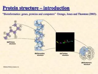
Structural Biologists • X-ray crystallography • NMR spectroscopy • High-resolution electron microscopy • Theoretical modeling
Terminology Visualization: Exploring an existing molecular model. Modeling: Creating a new molecular model or changing the conformation or composition of an existing model.
Visualization software you have used • PyMOL? • Chimera? • Jmol? • Something else? • RasMol? • Chime? • Protein Explorer? Obsolete
Workshop Overview Obtaining 3D Macromolecular Models Understanding The Models Sharing Structure-Function Information
Workshop Overview Obtaining 3D Macromolecular Models Published Empirical (Crystallographic or NMR) Homology Models (Theoretical Models Are Grossly Inaccurate) Understanding The Models Sharing Structure-Function Information Martz – May 2010
Workshop Overview Obtaining 3D Macromolecular Models Understanding The Models FirstGlance in Jmol ( + its links to Key Resources) Sharing Structure-Function Information Martz – May 2010
FirstGlance in Jmol ≠ Jmol! • Jmol (jmol.org) is a large international open-source project active since before 2000. • Lead developer since 2006 is Robert Hanson (St. Olaf College, Northfield MN). • Jmol is no easier to use than PyMOL, etc. • FirstGlance in Jmol (firstglance.jmol.org) is a user interface that makes Jmol’s power accessible to a wide range of users. • First available in 2006, it is developed by Eric Martz, following his experience with Protein Explorer. It is used in 3D View links in Nature Structure.
Workshop Overview Obtaining 3D Macromolecular Models Understanding The Models Sharing Structure-Function Information: Custom Molecular Scenes without Tears Polyview-3D: publication quality; animations for Powerpoint Proteopedia: a wiki; rotating molecules; on-line Martz – May 2010
Gal 4 Transcriptional Regulator Protein. DNA Double Helix (Chain / Chain). Lysine 18 sits in DNA Major Groove recognizing CGG. Martz – May 2010 – 1d66
Animation Created by Polyview-3D (using PyMOL) Martz – May 2010 – 1d66