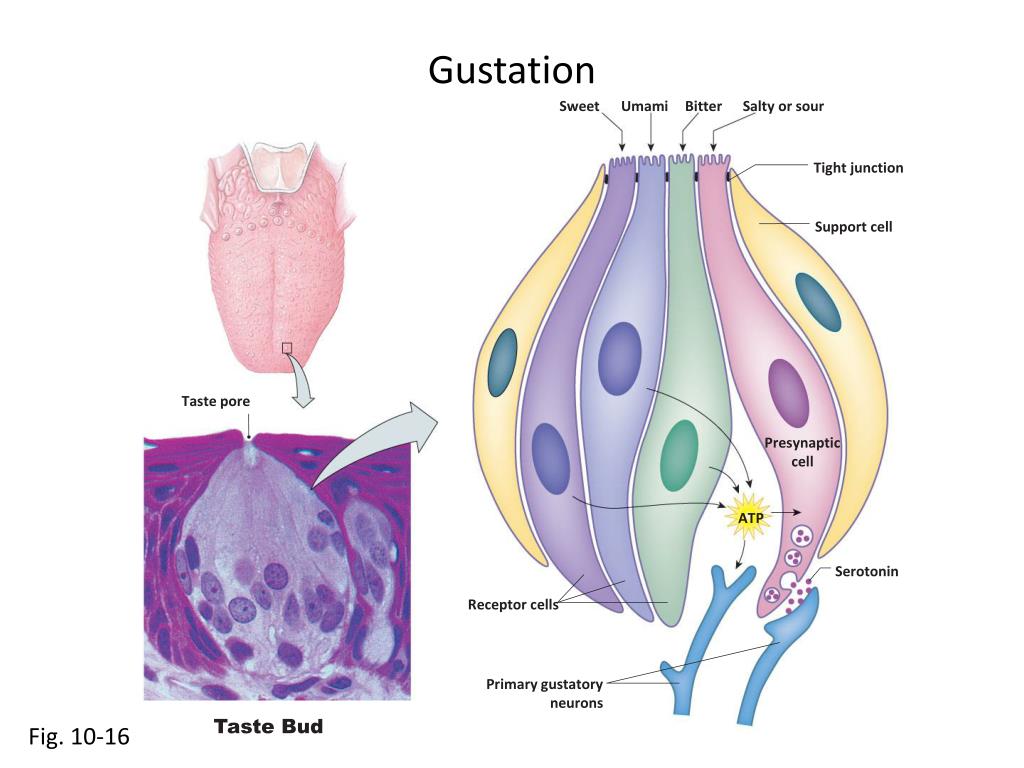
Gustation Sweet Umami Bitter Salty or sour Tight junction Support cell Taste pore Presynapticcell ATP Serotonin Receptor cells Primary gustatoryneurons Taste Bud Fig. 10-16
Taste Transduction Salt Sour Sweet, umami,or bitter ligand H+ Na+ 1 Gustducin 1 1 GPCR 2 2 2 Na+ H+ Signaltransduction Celldepolarizes ? ? Ca2+ Ca2+ 3 3 Ca2+ Ca2+ 3 Ca2+ Ca2+ ? ATP Serotonin 4 4 4 Primary gustatoryneurons 5 5 5 Fig. 10-17
Receptor A Receptor B Receptor C Receptor D
Olfaction Olfactorybulb Olfactorytract Olfactoryepithelium Fig. 10-14
Olfaction Brain Olfactory bulb Mitral cells Glomeruli To limbic system and cerebral cortex Bone Cilia Olfactory receptors
Olfactory Receptors Developing olfactory cell Olfactory receptor cell Supporting cell Olfactory cilia Mucus layer Fig. 10-14
Touch Receptors Merkel receptors Meissner’s corpuscle Hair Free nerveending Hair root Sensory nerves Paciniancorpuscle Ruffinicorpuscle Fig. 10-11
Hair Cells (a) At rest (b) Excitation (c) Inhibition Tip link Channels closed.Less cation entryhyperpolarizes cell. Somechannelsopen More channelsopen.Cation entrydepolarizescell. Stereocilium Hair cell Primarysensoryneuron Action potentials Action potentials increase No action potentials mV Action potentials inprimary sensory neuron Time 0 mV –30 Release Release Membrane potentialof hair cell Inhibition closesion channels Excitation opension channels Fig. 10-22
Hair Cells
Anatomy of the Ear EXTERNAL EAR MIDDLE EAR INNER EAR Semicircularcanals Nerves Cochlea Vestibularappartus Fig. 10-18
Vestibular Apparatus SEMICIRCULAR CANALS Superior Cristae withinampulla Horizontal Posterior Cochlea Utricle OTOLITH ORGANS Saccule Maculae Fig. 10-25
Kinocilium Stereocilia Otolith organs Otoliths Gelatinous layer Hair cells Supporting cells Similar toFig. 10-25 Sensory nerve fibers
Otolith Movement
Vestibular Apparatus SEMICIRCULAR CANALS Superior Cristae withinampulla Horizontal Posterior Cochlea Utricle OTOLITH ORGANS Saccule Maculae Fig. 10-25
Orientation of Semicircular Canals Vestibularapparatus Superior canal(nod for “yes”) Posterior canal(head tilt) Left right Horizontal canal(shake headfor “no”)
Endolymph Movement Cupula Bone Endolymph Hair cells Bone Direction of rotation of the head
Anatomy of the Ear EXTERNAL EAR MIDDLE EAR INNER EAR The pinnadirects soundwaves intothe ear Ovalwindow Malleus Incus Nerves Stapes Cochlea Earcanal Tympanicmembrane Roundwindow Topharynx Eustachiantube Fig. 10-18
Anatomy of the Inner Ear Ovalwindow Vestibularduct Cochlearduct Organ ofCorti Saccule Cochlea Uncoiled Helicotrema Roundwindow Tympanicduct Basilarmembrane
Anatomy of the Inner Ear Bony cochlear wall Vestibular duct Cochlear duct Tectorial membrane Organ of Corti Cochlear nerve transmitsaction potentials fromthe hair cells to theauditory cortex. Basilarmembrane Tympanicduct
Transmission of Sound Cochlear nerve Incus Ovalwindow Malleus Stapes Ear canal 5 Vestibular duct(perilymph) 3 Cochlear duct(endolymph) 2 6 4 1 Tympanic duct(perilymph) Tympanicmembrane Roundwindow
Transmission of Sound The movement of the tectorialmembrane moves the cilia onthe hair cells. Fluid wave Cochlearduct Tectorialmembrane Haircell Tympanicduct Nerve fibers ofcochlear nerve Basilar membrane
Anatomy of the Eye Zonular fibers Lens Optic disk(blind spot) Canal ofSchlemm Central retinalartery and vein Aqueoushumor Cornea Optic nerve Pupil Fovea Iris Vitreous chamber Retina Ciliary muscle Sclera is connective tissue. Fig. 10-30
Anatomy of the Eye Horizontalcell Amacrinecell Light Ganglioncell Cone (color vision) Neurons where signalsfrom rods and conesare integrated Bipolarcell Rod (monochromatic vision) Fig. 10-37
Anatomy of the Eye Pigment epitheliumof retina absorbsexcess light. Rod Bipolar neuron Light Cone Fovea Ganglion cell Fig. 10-37
Focusing Mechanism Light Lens Retina Optic nerve Fixationpoint Macula Fovea Fig. 10-37
Focusing Mechanism Ciliary muscle Lens Cornea Ligaments Iris Fig. 10-34
Focusing Mechanism Ciliary musclecontracted Ciliary musclerelaxed Lens rounded Lens flattened Cornea Ligamentsslacken Ligamentspulled tight Fig. 10-34
Photoreceptor Structure OUTER SEGMENT Disks Disks Connectingstalks INNER SEGMENT Mitochondria Rhodopsinmolecule Cone Rods Retinal Opsin SYNAPTIC TERMINAL Bipolar cell LIGHT Fig. 10-39
Photoreceptor Sensitivity Fig. 10-39
Integration of Signals Pigmentepithelium To opticnerve Bipolarcell Rod Ganglioncell Fig. 10-37