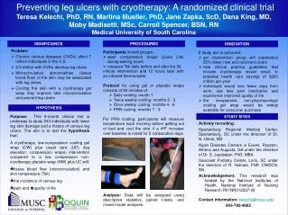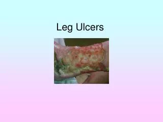
Leg Ulcers
Leg Ulcers. Dr. Raghunandan. Over view . Definition Problem – How big is it ? Types Pathophysiology of venous , arterial , diabetic ulcers Assessment / Evaluations Treatment options – Dressing agents , surgical options . Ulcers.

Leg Ulcers
E N D
Presentation Transcript
Leg Ulcers Dr. Raghunandan
Over view • Definition • Problem – How big is it ? • Types • Pathophysiology of venous , arterial , diabetic ulcers • Assessment / Evaluations • Treatment options – Dressing agents , surgical options
Ulcers • An ulcer is defined as an area of discontinuation of the surface epithelium. • A leg ulcer is a discontinuity of the squamous epithelium of the skin usually around the ankle or on the foot
Chronic Ulcer • A chronic leg ulcer is more difficult to define but many people consider ulceration of more than 4-6 weeks duration as being chronic. • Chronic ulcers results when sequel of repair is disturbed at one or more stages of inflammation , proliferation , re epithelialization ,remodelling • Staph aureus , Strep pyogens , Strep fecalis , E coli are common organisms colonizing the ulcers
Incidence • 12/10000 - Irish data • 2-4% of the population at any given time will have ulcers due to venous disease • 0.06-0.20% of the total population • Average age of patients 70 years – increased as more people are living longer • Women are twice likely to be affected than men .
Diabetes – Facts • 16 million diabetics • 15% develop foot related problems • 30% all hospitalizations due to foot related problems • 50000 amputations • 50% develop contra lateral foot problems and 50% again will have amputations • 3 year mortality is approximately 50% .
Etiology • Venous • Arterial • Mixed –arterial and venous • Neuropathic –Diabetes • Connective tissue disorders- vasculitis • Infective – tuberculosis. • Malignancy • Trauma
Venous ulcers • Ankle pressure at ankle when standing is 125 cms H2O but on walking the action of calf muscles surrounding the vein pushes the blood out of the leg and reduces the pressures to about 40 cms of H2O
Venous ulcers • Reflux • Superficial or deep veins • Combination • Obstructive • Primary varicose veins • Secondary veins venous hypertension
Venous hypertension • Increased pressure at ankle • Swelling of the tissues • widening endothelial gap junctions • Sequestration of the RBCs, WBCs , Proteins
Post thrombotic events • Obstruction • Valves get damaged during healing process • Chronic venous insufficiency • Poor venous return
Venous hypertension • Fibrin cuff theory • Increased venous pressure • Loss of plasma proteins • Fibrinogen forms a cuff around the capillaries • Fibrin cuff interferes with the exchange of oxygen • Tissue breaks down
Venous hypertension • Leukocyte migration theory • White cells migrate into the interstitial tissue • break down of the WBCs lead to the cytokines and proteases release . • Loss of tissue integrity
Arterial occlussion • Indicate the presence of severe occlusive disease . Atherosclerosis , vasospasm , inflammatory vascular disease / • loss of nutrients and oxygen lead to tissue break down • arterial ulcers are common in the feet , head of the 1st and 5th metatarsals .
Diabetes • Hyper glycemia leads to increase in glucose content in the tissues which binds to proteins leading to cellular damage • Increase sorbitol and fructose in cells leads to accumulation of water in the cells • Increased sorbitol leads to decreased myoinositol in cells also postulated for the cellular damge • Neutrophil dysfunction and phagocytosis
Diabetic ulcers • Vision loss • Shoe trauma / Thermal injury • Charcots foot ( neuro osteoarthropathy) • Six times more incidence of PAOD than the rest of the population
Diabetes • Summary • Ischemia • Neuropathy • Infections
Other causes • Malignancy • Trauma – osteomyelitits • Infections – TB . • CTD – vasculitis
Why assessment • Pre requisite for the effective leg ulcer management • Minimizes improper use of treatment • Reduces the risk of long term ulcerations • Facilitates early detection of life or limb threatening problems • For developing strategies to limit the recurrences
Assessment • Allows • Etiology of ulcers • Local or general factors that may cause a delay in healing • Social circumstances and optimum setting for care
Assessment • Falls into • Medical history • Physical examination • non invasive evaluations • Invasive evaluations
Ulcer examination • Site • Size • Shape • number • floor • edge / margin • Base • surrounding skin • Examination of the arterial . Venous , lymphatics , neurological system • evaluation of the nutritional status and underlying medical conditions that prolong wound healing
Ulcer evaluations • highest ankle pressure ABI = ----------------------------- Highest brachial pressure • For screening of the arterial disease • For compression therapy • For monitoring purposes
Ulcer evaluations • FBC,ESR,Renal & Liver functions • Wound swab and qualitative cultures • Duplex studies of the venous system • Connective disease profile • X-ray of the long bones • Angiography • Biopsy of the ulcers ( Marjolins ulcers)
Ideal dressing agent • Protect from bacterial invasion • maintain optimum humidity • absorb serum from wound site • protect granulation tissue • reduce pain
Goals for therapy • Debridement – Mechanical / surgical / biological / enzymatic • Off loading foot wear . • Antibiotics • Appropriate wound care .
Dressing agents • No role for • Hydrogen peroxide • Boric acid • EUSOL • Dakin solution (hypochlorite ) • Iodine As they are toxic to the tissues
Dressing agents • Poly urethane films • transmit water vapour , oxygen , carbon di oxide • non absorbent • useful for healing wounds with minimal drainage • Foams and Hydrocolloids • Permeable , easy to apply , minimum re injury when removing the dressings • 60-95% water content maintains the moist atmosphere
Dressing agents • Alginates • Sea weed preparation • absorb exudates • useful for exudative wounds • Cultured keratinocytes • Cells are cultured and transferred to petroleum gauze • labour intense and expensive
Growth factors and wound healing • They are poly peptides , stimulate wound healing , promote chemotaxis , miotgenesis of fibroblasts and smooth muscle cells • Plate let derived growth factor , Insulin like growth factor , epidermal growth factor , fibroblast growth factor , transforming growth factor 1
Compression therapy • Developed by the Charing cross group • Different sizes for various ankle diameters • Main stay of the venous disease • Prevention and treatment • <0.8 ABI will need further assessment • improves healing rate compared to no compression therapy • Multi layer better than single layer • higher the pressure better the healing rate
Profore • Multiple layer bandage for the venous hypertension • Padding , crepe , light compression ,high compression layers • 0.6 – 0.7 ABI – use Profore lite • ABI <0.5 contraindication for the compression therapy
Management issues • Nutrition-proteins , zinc , vitamin c • Pain management • Change of dressings • Removal of slough- hydrogels , varidase • decrease the bacterial load – iodoflex • Reduction of exudates- alginates • Odour – iodoflex, silver , metronidazole • Eczema- steroids
Role of antibiotics • Bacteria can secondarily colonize the wound and general tendency is to over treat . • Not necessarily indicate infection • wound bacteria may be transient and may not be detected on random swabs • Fever /erythema /swelling / increased pain / leucocytosis
Management issues • Long term use of compression therapy is useful in preventing the recurrences • Below knee stockings are as good as above knee stockings • Replace every 6 months • To be used for the day time and foot care at night • keep foot end elevated.
Management issues • Education – • avoid standing for long duration • Walking • to keep physically active • care of foot • 20% chances of recurrences
Surgery for lower limb ulcers • Venous . • Varicose vein – SFJ / SPJ ligation , GSV stripping , Avulsion of varicosities . • Sub fascial perforator surgery • Deep vein reconstruction • Arterial • Angioplasty • Bypass procedures