Download
1 / 35
460 likes | 915 Vues
Necropsy-examination of an animal after it has died. Necropsy: Fun at the Beach. Terms. Pathology—study of disease Gross pathology—pathologic changes seen with naked eye Histopathology—clinical changes seen with a microscope Lesions—pathologic changes

E N D
Terms Pathology—study of disease • Gross pathology—pathologic changes seen with naked eye • Histopathology—clinical changes seen with a microscope • Lesions—pathologic changes • Pathogenesis—the sequence of events of the disease (dog was bitten → bacteria entered wound → elevated temperature → pus formed → etc)
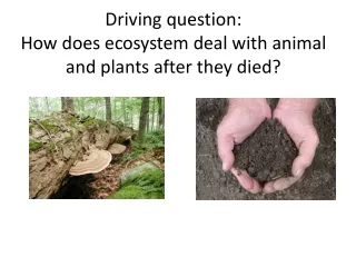
Reasons for Necropsy • Determine cause of death • Determine accuracy of clinical diagnosis • Evaluate effects of therapy • In herds, 1 or more animals may be sacrificed to determine the cause of disease or toxicity • Routine in pharmaceutical studies
Preliminary Steps for Necropsy • Obtain owners permission; also determine if owner wants remains for burial, etc • Correctly identify animal (species, breed, sex, age, ID tags or tattoos) • Perform necropsy ASAP after death; if delayed, refrigerate animal to delay autolysis; DON’T FREEZE
Necropsy Report • Location • Number of lesions • Color of abnormalities (dark red, black, etc) • Size of lesions (cm or weight) • Shape of lesion (round, flat, oval) • Distribution • Consistency (hard, soft, firm) • Odor (sweet, sour, ammonia) • Final Report tense should be consistent • Report should be as specific as possible without giving a final diagnosis unless a test for the Dx was performed (i.e. rabies)
Necropsy: Protective Clothing • Plastic apron, lab coat, scrubs • latex or plastic disposable gloves • surgical mask if animal died from infectious disease spread by aerosolization • protective footwear
Necropsy: Tools • Knives • Scissors • Tissue forceps • Bone-cutting tools (pruning shears, hacksaw)
Toxicology Samples • blood (10-20 ml) • stomach contents and urine (50-100 ml) • blocks of liver, fat, kidney, and brain (5x5x10 cm; approx 200 g) • Samples
Necropsy Videos http://video.vet.cornell.edu/virtualvet/bovine/chapters1-4.html
Esophagus Normal Food Ulcerations Bleeding
Larynx normal necrosis ulceration
Trachea normal necrosis ulceration
Pericardial sac Click on picture to view video
Lungs Normal Bronchopheumonia Normal lung tissue -pink -spongy Lung abscess -liquid/“cottage cheese” like appearance
Airways and vessels Lung artery Lung airway
Lung lesions Emphysema -pops like bubble-wrap Consolidation -heavy; solid (no air) Lung worms Lung adhesions to ribs Pneumonia- darker lesions are more severe Abscesses
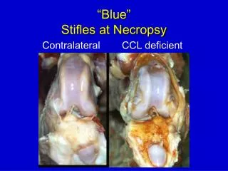
Heart Lesions Septal defects—connection between R and L sides Necrotic lesions Valve lesions
Liver lesions Liver abscesses Liver flukes Fatty liver Fractured liver—due to blunt trauma; (knife cuts smooth)