Advanced Techniques in DTI for Sensorimotor Cortex Analysis and Motion Correction
This presentation explores advanced Diffusion Tensor Imaging (DTI) techniques and their application in analyzing the sensorimotor cortex. Key topics include segmented sampling, various motion correction methods, and fiber orientation estimation using Constrained Spherical Deconvolution (CSD). We highlight the efficacy of fMRI-based Regions of Interest (ROIs) compared to traditional methods, emphasizing approaches that enhance the accuracy of fiber tracking and motion correction. Techniques such as angular resolution, diffusion-weighted scans, and simulation methods for head movement correction are also discussed.

Advanced Techniques in DTI for Sensorimotor Cortex Analysis and Motion Correction
E N D
Presentation Transcript
David Schaeffer (University of Georgia) Lauren Libero (University of Alabama at Birmingham) Sara Levens, Ph.D. (University of Pittsburgh) DTI ModuleMNTP 2011 Instructor Kwan-Jin Jung, Ph.D. (Carnegie Mellon University) Technical Assistant NidhiKohli (Carnegie Mellon University)
Effects of • Segmented sampling • Motion correction • Fiber orientation estimation method • fMRI based ROIs vs. drawing ROIs • Anatomical separation of sensorimotor cortex Learning Objectives
Diffusion encoding gradient direction • Vector table (x, y, z components) • Angular resolution • Diffusion-weighting (b-values) • Duration & amplitude • s/mm² • b0 = 0 s/mm² • No diffusion gradient Terminology
Segmented sampling • Complementary diffusion encoding directions • 64 (A) - 10 min • 64 (B) - 10 min • 128 (A + B) - 20 min • Useful for special populations Method of Acquisition
How to correct: 1. Estimate the motion 2.Rotate image and vector table accordingly Motion Correction Intended Collected Head correction WRONG Head & vector table correction CORRECT
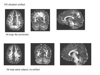
No correction • No vector rotation • Interpolation • Estimates how much you rotate vector table • Based on distributed b0 images – “real motion” Motion Correction 6 6 Rotation (degrees) 3 3 Rotation (degrees) 0 0 -3 -3 -6 -6 Time Time BEFORE AFTER
Simulation method • Collect two diffusion scans • 6 direction scan (low b-value) • Why? – Fast (little time for motion) • Edges of brain are clearly defined • 6 or more direction scan (higher b-value) • Assume no motion on scan 1, then simulate what higher b-value volume should look like Motion Correction
Find D (diffusion tensor) S=S0e-bD Find S (simulated high b-value) S=S0e-bD Low b-value (b=800 s/mm²) DWI (scan 1) Assume no motion Co-register volumes (estimating motion) High b-value (b=2000 s/mm²) DWI (scan 2) Rotate vector table
Tensor • Performs well for straight tracts (like motor) • Performs poorly for crossing and branching fibers (like Genu) • Constrained Spherical Deconvolution (CSD) • Better for detecting branching and crossing fibers Fiber orientation estimation method (Tournier et al., 2007)
Csd Vs. Tensor Genu CSD Genu Tensor
Manually draw ROIs • Using fMRI • Collect fMRI data – find center of activation (x, y, z) • Matrix transformation • Convert from fMRI coordinates into DWI native space DrawingROIs
Finger closing fMRI results as ROI • Separation of sensory and motor areas • Clustering – fiber end-point distribution Segmenting Sensorimotor Central Sulcus
Sampling schemes can be advantageously altered for use with special populations Simulation is a promising method for more accurate motion correction CSD Fiber tracking is most appropriate for resolving fiber crossings summary
fMRI-based ROIs can be used to track fibers from areas of activation DTI can be used as a tool to segment brain areas that are not separable based on diffuse fMRI activation maps Summary
Dr. Kwan-Jin Jung • NidhiKohli • MNTP Leaders: Dr. Eddy & Dr. Kim • MTNP Trainees & Participants • DTI Trainees 2009 & 2010 • Funding: • NIH grants: R90DA023420 and T90DA022761 Acknowledgments
motion correction No correction Interpolation Simulation Motion