Download
1 / 37
500 likes | 1.36k Vues
Diagnostic Evaluation in Child Abuse. Robert Benowicz, MS4 OHSU. Introduction. Definitions/Epidemiology Clinical Manifestations Skeletal Trauma Soft Tissue/Visceral Trauma Head Trauma Diagnostic Evaluation Management/Psychosocial Considerations. Definitions.

E N D
Diagnostic Evaluation in Child Abuse Robert Benowicz, MS4 OHSU
Introduction • Definitions/Epidemiology • Clinical Manifestations • Skeletal Trauma • Soft Tissue/Visceral Trauma • Head Trauma • Diagnostic Evaluation • Management/Psychosocial Considerations
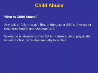
Definitions • Child Abuse Prevention and Treatment Act (CAPTA) • Any recent act or failure to act that results in death, serious physical or emotional harm, sexual abuse, or exploitation; or imminent risk of serious harm • Involves a child under the age of 18 and his/her parent or caretaker • Types of Abuse • Physical Abuse • Sexual Abuse • Emotional Abuse • Child Neglect
Epidemiology • Annual Incidence 15-42 cases per 1000 children • Greater than 1 million children are victims of abuse/neglect per year • Over 1200 children die annually as a result of abuse with almost half of these cases occurring in ages less than 12 months • Minorities have higher rates of reported abuse than do whites • But minorities tend to be evaluated and reported for suspected abuse more frequently than whites
Epidemiology • Usual Suspects • Fathers, mother’s boyfriends, female babysitters, mothers • Risk Factors • Young/single parents, unstable families, lower education levels, drug/EtOH abuse, psych illnesses, parents/caregivers whom were abused themselves, unrealistic expectations of their children, poor coping skills, financial stresses • Victims of Abuse • Age (majority of cases are less than 12 mos) • Past Hx of Abuse • Disabilities (physical, learning, speech/language) • Psychiatric (hyperactive, conduct disorders) • Medical Conditions (chronic illnesses, prematurity/low birth wt)
Clinical Manifestations • Skeletal Trauma • Eg. Fractures • Skin Trauma • Eg. Bruises, burns • Visceral Trauma • Eg. Splenic lacerations, hepatic contusions, GI perforation • Head Trauma • Eg. Scalp injury, skull fracture, intracranial injury, facial injury
Skeletal Trauma • Patterns • Most common fx involve skull, long bones, and ribs • Nonaccidental fx most common in children under 3 yrs of age (infants especially) • Classic Metaphyseal Lesions (CML), skull and rib fx most common in infancy (<12 mos) • Long bone shaft fx are more common in toddlers and are the most prevalent in all cases of child abuse
Long Bone Fractures • A-longitudinal • B-transverse • C-oblique • D-spiral • E-impacted • F-comminuted • G-greenstick • H-bowing • I-torus
Diaphyseal Fractures • Diaphyseal vs Metaphyseal • Four times more common • Femur, Humerus, Tibia • most common sites • Spiral and transverse most prevalent, but greenstick fx (right) also occur • Ambulatory vs Non-ambulatory • Amb toddlers have higher rate of accidental fx • Suspect abuse in non-ambulatory infants
Metaphyseal Fractures • Classic Metaphyseal Lesion (CML) • Aka “corner fracture” or “bucket-handle fracture” • Triangular or disk-like fragment of the metaphysis • Occur when extremity is pulled hard or twisted and can occur during violent shaking • Typically asymptomatic
Rib Fractures • Uncommon but frequently indicative of child abuse • 5-27% of all skeletal injuries in one study • Can also occur after serious accidental injuries, birth trauma, or 2° to bone fragility • Anterior-Posterior compression • 94% of abused infants had posteriorly or laterally located fractures in same study • Bulloch et al. Pediatrics April 2000.
Other Fractures • Spinal • most commonly L1-2 • Cervical injury w/ violent shaking of head/neck • Very few cases reported • Hands/Feet • Torus/buckle fx • Sternum, Scapula, Pelvis • fractures not highly specific for abuse but outside of high energy traumatic injury suspicion should be high • Multiple Fractures (right) • High suspicion of abuse, especially if the fx are in various stages of healing • Levin et al. Ped Radiol (2003) • Hecther et al. Ped Radiol (2002)
Great Imitators of Skeletal Trauma • Skeletal Fractures • Accidental trauma • Eg. Toddlers Fx (top right) • Normal variants • Birth trauma • Pathologic fractures • Metabolic bone disease • Rickets • Neoplasm • Skeletal Dysplasia • Osteogenesis Imperfecta (bottom right) • Periosteal Reaction • Infection • Osteomyelitis, Congenital syphilis • Drug toxicity • Vit A, MTX, Prostaglandin
Skin Trauma • Bruises • Most common type of injury related to abuse • Orofacial injuries • Racoon eyes • Traumatic alopecia • Ruptured TM’s • OP gonorrhea or syphilis • Noninflicted bruises on bony prominences whereas inflicted bruises are more central • Multiple bruises in clusters raises suspicion of abuse • Immobile infants with specific bruising patterns should raise suspicion
Skin Trauma • Burns • Occur in 6-20% of physically abused children • Specific patterns can be highly suggestive of abuse • Types • Brands/contact burns • Cigarette burns • Immersion burns (most common) • Caustic material burns • Delay in seeking medical attention for abusive burns
Great Imitators of Skin Trauma • Bruises • Bleeding disorders • ITP, hemophilia (VIII/IX), vonWIllebrand disease • Vasculitis • HSP • Mongolian spots • Burns • Phytophotodermatitis (top right) • Impetigo • Cultural Practices • Cupping (bottom right) • Coining • Salting
Visceral Trauma • Typically occur in setting of high-energy trauma • Suspect inflicted injury if there is no such history • Thoracic trauma • Esophageal perforation, pulmonary lacerations/contusions, chylothorax • Cardiac trauma • Dysrhythymias, contusions, traumatic VSD • Abdominal trauma • Liver (most common), splenic, pancreatic, GI tract (perf vs hematoma) • Urinary tract trauma • Renal, ureteral, bladder
Head Trauma • Initial assessment • Bradycardia, apnea/brachypnea, hypothermia, poor motor tone, nystagmus, seizures, bulging fontanel may all be present • Numerous retinal hemorrhages at multiple layers of the retina is highly suggestive of shaken baby syndrome • Optho consult if available • Cutaneous lesions are not as specific as retinal hemorrhage but can hint at further abuse
Head Trauma • Levels of damage • Scalp hematomas • Skull fractures (top right) • Epidural hemorrhages • Focal subdural hemorrhages (bottom right) • Brain contusion/laceration • Distant sites • Basilar skull fractures • Retinal hemorrhages often present • Brainstem compression • Coma and/or death
Great Imitators of Head Trauma • Non-accidental Head Injury Mimics • Benign Infantile enlargement of the subarachnoid space • Symmetric, absence of assoc. lesions, and absence of blood products • Diastatic sutural injury vs. sutural splitting/widening from increased ICP • Hemorrhage due to DIC, infection, anticoagulant therapy • Accidental trauma • Falls from less than 3 feet rarely produce severe head injury • Edema due to smoke inhalation, drowning, or circulatory collapse • Demaerel et al. Eur Radiol (2002)
Initial Evaluation • Medical Evaluation • History • Physical Examination • Observation of Parent’s Behavior • Laboratory Evaluation • Bleeding studies • Urinalysis • Chemistries • Toxicology screen • Radiographic Evaluation
Diagnostic Evaluation • Skeletal Trauma • Skeletal survey vs. scintigraphy • Methods of global imaging in suspected child abuse • Orthogonal radiographs • Bone tenderness, swelling, deformity, limited ROM • Recommendations • 0-12 mos. (reqd) • Skeletal survey • F/U survey at 2 weeks • 12 mos to 2 yrs (reqd) • Skeletal survey or scintigraphy • 2 to 5 yrs • Skeletal survey or scintigraphy in cases where abuse is strongly suspected • 5 yrs and older • Radiographs of individual sites of injury suspected on clinical grounds • Kleinman, PK. Diagnostic Imagingof Child Abuse, 2nd ed.
Diagnostic Evaluation • Visceral Trauma • Thoracic Injury • Orthogonal CXR and C-spine are initial tests of choice • Followed by CT if patient is stable and further injury is suspected • Esophageal injury may necessitate contrast esophagography or CT w/ oral contrast • Abdominal Injury • Plain flat/upright radiographs are obtained as part of the initial evaluation • Further testing warranted based on vital signs, physical exam findings, and lab results • Upright or decubitus views if bowel perforation suspected • Indications for CT • History/PE suggestive of significant abd injury, hematuria, decreased HCT, elevated AST/ALT, unaccountable fluid loss/requirements • Ultrasound is specific but not sensitive and is avoided in favor of CT • Barium upper GI series w/ small bowel follow-through
Diagnostic Evaluation • Head Trauma • There is a major debate over the preferred methods of radiographic evaluation of non-accidental head injury (NAHI) in the literature • Skull fractures • Skull x-rays are a part of the standard skeletal survey • Do not add much information over more advanced neuroimaging especially if head trauma is suspected upon initial evaluation • Suspected fractures of the cranial vault, however, are better seen by x-ray than CT • Intracranial injury • Nonenhanced CT is the imaging tool of choice • Some camps (Demaerel et al. and Stoodley et al.) believe that further investigation w/ MRI is also warranted • Other camps (McHugh et al.) believe that even discretion with head CT is necessary (see next slide) • MRI indicated when NAHI suspected but CT is normal • Can detect very small hematomas or subtle extra-axial fluid collections that might be otherwise missed • Also good for dating intracranial injury
Diagnostic Evaluation • Indications • Known skull fx • Altered mental status (see coma scale) • Focal neuro deficits • Signs of basilar skull fx • Retinal hemorrhage • Seizure • Palpable skull depression • Age • <2 yrs of age,<12 mos, and <6 mos have been proposed as cutoffs • Infants and toddlers may have fewer clinical findings and thus the CT threshold should be lower
Management • Suspected Abuse • Multidisciplinary team • Social worker, case management, nursing staff, other physicians • Hospitalization • All medical issues are addressed first • Child’s safety is addressed once patient is medically stable • Protective environment needed until Child Protective Services (CPS) can do an official evaluation • Children re-released to a caregiver have a 50% chance of being subjected to abuse again and a 10% mortality rate • Talking with parents • Mandatory reporting • Documentation
Psychosocial/Medicolegal • Talking with Parents • Inform parents why investigation is taking place • Safety and well-being of child • Required by law • Explanation of the CPS process • Mandatory Reporting • According to Oregon Revised Statute 419B.010, "Any public or private official having reasonable cause to believe that any child with whom the official comes in contact has suffered abuse, or that any person with whom the official comes in contact has abused a child shall immediately report or cause a report to be made . . ." Those "public or private officials" include: • Among others • Physician, including any intern or resident • Licensed practical or registered nurse • http://www.oregon.gov/DHS/children/abuse/cps/report.shtml
References • Kleinman, PK. Diagnostic Imaging of Child Abuse, 2nd ed. (1998). • Bulloch et al. “Cause and Clinical Characteristics of Rib Fractures in Infants.” Pediatrics Vol 105 No. 4 April 2000. • Hechter et al. “Sternal Fractures as a Manifestation of Abusive Injury in Children.” PediatricRadiology 32: 902-906, (2002). • Levin et al. “Thoracolumbar Fracture with Listhesis—An Uncommon manifestation of Child Abuse.” Pediatric Radiology 33: 305-310, (2003). • Demaerel et al. “Cranial Imaging in Child Abuse.” European Radiology 12: 849-857, (2002). • Carty et al. “Non-accidental Injury: A Retrospective Analysis of a Large Cohort.” EuropeanRadiology 12: 2919-2925, (2002). • Stoodley et al. “Neuroradiological Aspects of Subdural Hemorrhages.” Arch Dis Child 90: 947-951, (2005). • Stoodley et al. “Apnea and Brain Swelling in Non-accidental Head Injury.” Arch Dis Child 88: 472-476, (2003). • McHugh, K. “Neuroimaging in non-accidental Head Injury: If, when, why, and how” Clinical Radiology 60(1):22-30 Jan 2005. • http://www.uptodate.com