Download

1 / 18
410 likes | 2.61k Vues
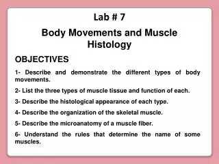
Muscle Histology. Cross Section of a Skeletal Muscle . Blue = epimysium Green = perimysium Yellow = endomysium surrounding muscle fiber. Pointer is on the perimysium surrounding a muscle fascicle. Skeletal Muscle Cross Section Slide.

E N D
Cross Section of a Skeletal Muscle • Blue = epimysium • Green = perimysium • Yellow = endomysium surrounding muscle fiber
Pointer is on the perimysium surrounding a muscle fascicle Skeletal Muscle Cross Section Slide Epimysium would be here surrounding the entire muscle organ (but it is not visible on the slide)
Skeletal Muscle Cross Section Slide Muscle fascicles can be a variety of shapes!
Skeletal Muscle Cross Section Slide Pointer is on the perimysium surrounding a muscle fascicle black arrows are pointing at individual skeletal muscle fibers
Multinucleated Skeletal Muscle Fibers The small purple dots are the nuclei of a skeletal muscle fiber. Notice that each fiber has more than 1 nucleus?
Skeletal Muscle Long Section • Each bracket is on one Skeletal Muscle Fiber (cell)
Skeletal Muscle: Striations Skeletal muscle fibers are clearly striated with dark A-bands and light I-bands
Skeletal Muscle Long Section:Multinucleated Skeletal muscle fibers are multinucleated (they have more than 1 nucleus per cell)
Cardiac Muscle • Intercalated discs • Striations • Nuclei
black arrows are pointing at intercalated discs (only found on cardiac muscle)
Smooth Muscle • Nuclei Smooth muscle cells Smooth muscle cells have a spindle shape and only one nucleus per cell with NO striations!
Motor Unit = a somatic motor neuron + skeletal muscle fibers it synapses with
Motor Unit Slide • Green = Neuromuscular Junction • Yellow = Axon of a somatic motor neuron • White = skeletal muscle fiber
Motor Unit Slide Pointer is on the axon of a somatic motor neuron Green arrow is on a neuromuscular junction