Download
1 / 25
331 likes | 947 Vues
ANPS 19. Skeletal Muscle Histology. Dr. Tompkins Department of Neurological Sciences Given D408 john.tompkins@uvm.edu. Reading: Anatomy & Physiology: An Integrative approach, McKinley et al., Chapter 10, Muscle Tissue, pages 331-340 (Sections 10.1-10.2). Muscle Tissue.

E N D
ANPS 19 Skeletal Muscle Histology Dr. Tompkins Department of Neurological Sciences Given D408 john.tompkins@uvm.edu Reading: Anatomy & Physiology: An Integrative approach, McKinley et al., Chapter 10, Muscle Tissue, pages 331-340 (Sections 10.1-10.2)
Muscle Tissue • Muscle tissue distributed almost everywhere • Some functions of muscular tissue • Produces skeletal movement • Propels food we eat along gastrointestinal tract • Expels waste we produce • Changes amount of air that enters the lung • Pumps the blood to body tissues (all these functions accomplished by different types of muscle tissue)
Muscle Tissue • Three types of muscle tissue: • Skeletal muscle, cardiac muscle, smooth muscle • In contrast to cardiac and smooth muscle, skeletal muscle is under voluntary control • Composes 40-50% of weight of the adult • 700 skeletal muscles in the muscular system
Skeletal Muscle Functions • Produce skeletal movement • Maintain posture and body position • Support soft tissues • Storage and movement of materials sphincters - circular bands of skeletal muscle ensure the voluntary expulsion of feces and urine • Maintain body temperature
Characteristics of Skeletal Muscle Tissue • Excitable - responds to electrical stimulation (like neurons) • Conductive – action potential moves along membrane (like neurons) • Contracts • Elastic • Extendable
Structural Organization of Skeletal Muscle(Figure 10.1) • A skeletal muscle is composed of thousands of muscle cells, connective tissue, blood vessels & nerves. • Muscle cells (referred to as muscle fibers) are typically as long as the entire muscle. • Bundles of muscle fibers termed fascicles. Tendon (connects muscle to bone) Deep fascia (ensheathes functionally distinct muscles) Epimysium Skeletal muscle Artery • The epimysium, perimysium and endomysium are three concentric layers of connective tissue. • Provide protection, sites for blood vessel and nerve distribution, and are a means of attachment to the skeleton or other structures. Vein Perimysium Nerve Fascicle Endomysium Muscle fiber (This should look familiar to you. It’s similar to the structure of a nerve.)
Organization of connective tissues • Epimysium surrounds whole muscle • Perimysium sheathes bundles of muscle fibers: surrounds fascicles • Epimysium and perimysium contain blood vessels & nerves which branch into the muscle • Endomysium which covers individual muscle fibers • Tendons attach muscle to bone.
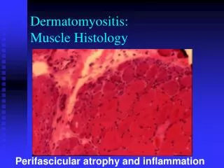
A cross sectional view of skeletal muscle. Where is the perimysium? Endomysium?
A high magnification view of skeletal muscle in cross section. C is for capillaries and P is for perimysium. Where is the endomysium? muscle fiber
Longitudinal section of skeletal muscle. Where is the endomysium?
Skeletal muscle fibers formed by fusion of myoblasts myo- or ( before a vowel ) my- from Greek mus muscle • These are the embryonic cells which fuse to form skeletal muscle fibers during development. Each contributes a nucleus to total number of nuclei. Myoblasts Unfused myoblasts termed “satellite cells”. These cells may be stimulated to differentiate if muscle is injured. Muscle fiber Myoblasts fuse to form a skeletal muscle fiber. Satellite cell Muscle fiber Nuclei Satellite cell muscle fiber is a multinucleated cell cytoplasm of muscle fiber termed sarcoplasm (Figure 10.2)
This is a diagram of a muscle fiber or a muscle cell showing the large number of myofibrils in the sarcoplasm
Inside a Skeletal Muscle Fiber • the endoplasmic reticulum of a muscle cell • surrounds bundles of contractile proteins (myofibrils) • contains the Ca2+ necessary for muscle contraction Sarcoplasmic reticulum Muscle Triad • part of the sarcoplasmic reticulum • a reservoir for Ca2+ ions • - 2 terminal cisternae + 1 T-tubule = 1 Triad Fascicle T-tubule Terminal cisternae Muscle fiber - plasma membrane of the muscle fiber - invaginations of this membrane termed T-tubules or transverse tubules Sarcolemma • - long cylindrical structure of fibers • - extend length of muscle fiber • - 80% of muscle fiber volume • - each fiber has hundreds to thousands Nucleus Myofibrils Myofilaments • protein filaments • thick and thin Nucleus Openings into T-tubules Sarcoplasm Sarcomere Nucleus • - functional unit of myofibril Mitochondrion (a) Skeletal muscle fiber (Figure 10.3 a)
Cross section of skeletal muscle fibers in which myofibrils can be seen.
Molecular Structure of Thick and Thin Filaments(Figure 10.4) Muscle fiber Myofilaments Myofibril - come in ‘thick’ and ‘thin’ Myosin molecule Heads Tail Actin binding site ATP and ATPase binding site Myosin heads (a) Thick filament Troponin Tropomyosin Ca2+ binding site F-actin G-actin Myosin binding site (b) Thin filament
Molecular Structure of Thick and Thin Filaments(Figure 10.4) Myosin molecule Heads Tail Actin binding site ATP and ATPase binding site Myosin heads • Thick filaments • assembled from bundles of protein molecules, myosin • each myosin protein has two intertwined strands • each strand has globular head and elongated tail • tails pointing toward center of thick filaments • heads pointing toward edges of thick filaments • head with a binding site for actin (on thin filaments) • head has site where ATP attaches and is split by ATPase
Molecular Structure of Thick and Thin Filaments(Figure 10.4) Troponin Tropomyosin • - globular protein attached to tropomyosin • - has binding site for Ca2+ • - together form troponin-tropomyosin complex • - twisted stringlike protein • - covers small bands of F-actin • - covers myosin binding sites in noncontracting muscle • Thin filaments • composed primarily of two strands of actin • strands twisted around one another • actin strands composed of spherical molecules, globular actin (G-actin) • connect to form a fibrous strand, filamentous actin (F-actin) • G-actin has myosin binding site - where myosin head attaches during contraction Troponin Tropomyosin Ca2+ binding site We will learn more about how these proteins work together in the next lecture. F-actin G-actin Myosin binding site
Organization of a Sarcomere • Myofibrils consist of repeating units called sarcomeres. • - number varies with length of myofibril. • - composed of overlapping thick and thin filaments • - delineated at both ends by Z discs • A band (DARK regions) • - central region of sarcomere • - contains entire thick filament • - contains partially overlapping thin filaments • - appears dark under a microscope Muscle fiber I band A band I band Sarcomeres Myofibril Z disc H zone Z disc Myofilaments M line Sarcomere (Figure 10.5 a) • Z discs • - containsspecialized proteins running perpendicular to myofilaments which anchor thin filaments • I bands (LIGHT regions) • - contains only thin filaments • - extend from both directions of Z disc • - appear light under a microscope • - disappear at maximal muscle contraction • H zone • - central portion of A band • - only thick filaments present; no thin filaments • - disappears during maximal muscle contraction • M line • - protein meshwork structure at center of H zone • - attachment site for thick filaments
A Sarcomere: The Functional Unit of Skeletal Muscle Contraction Sarcomere Z disc Z disc Thick filament Connectin (Titin) Thin filament Thin filament M line H zone I band A band I band (LIGHT) (DARK) (Figure 10.5 b)
Specific Banding Patterns in a Skeletal Muscle Fiber I – I band Z – Z disk A – A band N – nucleus These units aligned in a single fiber which contains many stacked myofibrils.
Structure of a Sarcomere Transverse sectional plane M line Thick filaments and accessory proteins H zone Thick filaments A band Thick filaments Thin filaments I band Thin filaments Connectin Z disc Thin filaments Connectin and accessory proteins (Figure 10.5 c)
Sarcomere Shortening Relaxed sarcomere Relaxed sarcomere Z disc Thick filament Z disc Connectin Thin filament Thin filament M line Z disc Z disc M line H zone H zone I band A band I band I band A band I band (a) Relaxed skeletal muscle Contraction Contraction M line Z disc Z disc Z disc Z disc M line A band A band Fully contracted sarcomere Fully contracted sarcomere (b) Fully contracted skeletal muscle (Figure 10.12)