Download
1 / 39
400 likes | 550 Vues
Delve into the intricacies of sleep physiology, EEG waves, NREM, and REM stages, uncovering the importance of each phase for overall health. Explore the role of neurotransmitters in sleep mechanisms and the impact of aging on sleep cycles. Gain insights into various sleep disorders and the significance of REM sleep for memory consolidation and brain function.

E N D
Dr. Eman El Eter Physiology of Sleep and EEG
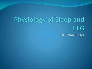
Objectives: • Difference between sleep & coma. • Why do we sleep? • Types of sleep: NREM & REM. • EEG waves. • Stages of NREM sleep. • Importance of REM sleep. • Sleep cycle and effect of age. • Sleep/awake cycle (Role of SCN). • Mechanism of sleep (centers/ neurotransmitters). • Sleep disorders.
Definition • Sleep is a state of loss of consciousness from which a subject can be aroused by appropriate stimuli. • Coma is a state of unconsciousness from which a subject cannot be aroused
Why do we sleep? • Restoration, or repair: • Waking life disrupts homeostasis • Sleep may conserve some energy • Protection with the circadian cycle • Circadian synthesis of hormones, …… • Consolidation of learning. • Remodelling of synaptic function
EEG waves • The frequencies of brain waves range from 0.5-500 Hz. • The most clinically relevant waves: • Alpha waves - 8-13 Hz • Beta waves - Greater than 13 Hz • Theta waves - 3.5-7.5 Hz • Delta waves - 3 Hz or less
Alpha waves • Seen in all age groups but are most common in adults. • Most marked in the parieto-occipital area. • Occur rhythmically on both sides of the head but are often slightly higher in amplitude on the nondominant side, especially in right-handed individuals • Occur with closed eyes , relaxation, wondering mind.
Alpha block:(Arousal response) • Alpha activity disappears normally with attention (eg, mental arithmetic, stress, opening eyes, any form of sensory stimulation). • Then become replaced with irregular low voltage activity. • Desynchronization as it represents breakup of synchronized neuronal activity. • An abnormal exception is alpha coma, most often caused by hypoxic-ischemic encephalopathy of destructive processes in the pons (eg, intracerebral hemorrhage). In alpha coma, alpha waves are distributed uniformly both anteriorly and posteriorly in patients who are unresponsive to stimuli
Beta waves • Seen in all age groups. • Small in amplitude , usually symmetric and more evident anteriorly. • > 13 Hz/sec
Theta waves • Normally seen during sleep at any age. • In awake adults, these waves are abnormal if they occur in excess. • Theta and delta waves are known collectively as slow waves.
Delta waves • Slow waves, have a frequency of ≤ 3Hz or less. • Normally seen in deep sleep in adults as well as in infants and children. • Delta waves are abnormal in the awake adult. • Often, have the largest amplitude of all waves. • Delta waves can be focal (local pathology) or diffuse (generalized dysfunction).
Sleep spindles • Spindles are groups of waves that occur during many sleep stages but especially in stage 2. • They have frequencies in the upper levels of alpha or lower levels of beta. • Lasting for a second or less, they increase in amplitude initially and then decrease slowly. The waveform resembles a spindle. • They usually are symmetric and are most obvious in the parasagittal regions.
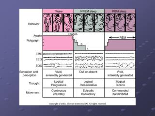
Types of sleep • 1. Slow-wave sleep (non-REM): • -75% of sleep time. • - restful. • - Decrease in vascular tone. • - Decrease in BP (10-30%) • - Decrease in Resp. rate. • - Decrease in BMR • It is not associated with rapid eye movement. • EEG: Theta + delta waves. • -If dreams occur they are not remembered as they are not consolidated in memory.
Types of sleep, continued,… • 2- Rapid Eye Movement Sleep (REM): • Is so called because of rapid eye moevement. • -Occur in episodes of 5-30 min, recurring every 90 min. • -Tiredness shortens the duration of each episode. • -As you become restful through the night, the duration of each episode increases. • -Active dreaming, remembered later.
REM, continued,…. • Decrease in muscle tone (due to excitation of reticular inhibitory centers). • HR & RR are irregular. • Rapid rolling movement of the eyes. • Erection of penis. • Engorgement of clitoris. • Twitches of facial & limb muscles. • More difficult to awake a person than in slow-wave sleep.
REM, continued,… • EEG: B-waves, indicating a high level of activity in the brain during REM (That is why it is called paradoxical sleep). PGO spikes stimulate the Inhibitory Reticular Area leading to Hypotonia: Exception: Respiratory + Eye muscles. In sleep apnea, respiratory muscles are inhibited.
Importance of REM sleep • 1. Expression of concerns in the sub-consciousness (Through dreams), • 2. Long-term chemical and structural changes that the brain need to make learning & memory possible.
Distribution of Sleep Stages • While NREM occupies 75-80% , it is interrupted by intervening REM sleep periods, every 90 minutes . • In a typical night of sleep , a young adult (1) first enters NREM sleep , passes through stages 1 , 2 , 3 and 4 , then • (2) goes into the first REM sleep episode. • This cycle is repeated at intervals of about 90 minutes throughout the 8 hours or so of a night sleep. • Therefore , there are 4-6 sleep cycles per night ( and 4-6 REM periods per night) • As the night goes on there is progressive reduction in stages 3 and 4 sleep and a progressive increase in REM sleep . REM sleep periods are shown in red (Hypnogram) In a young adult NREM occupies 75-80% of a night sleep time , & REM sleep occupies 20-25 % of the sleep time
Distribution of sleep stages in a typical night • Premature infants: • REM sleep occupies 80% of total sleep time. • Full term neonates: • 50% of sleep time is occupied by REM. • Aged/elderly: • The proportion of REM sleep falls rapidly and plateaus at about 25 % (20-69ys) until it falls further in old age . • Children have more sleep time and stage 4 than adults
Sleep/wakefulness rhythm • Periods of sleep and wakefulness alternate about once a day. • A circadian rhythm consist typically of 8h sleep and 16 h awake. • This rhythm is controlled by the biological clock function of suprachiasmatic (SCN) nucleus in the hypothalamus. • Within sleep portion of this circadian cycle NREM and REM sleep alternate.
Mechanism of Sleep NREM Sleep promoting systems: Preoptic area: VLPO and MNPO; are active during NREM sleep The neurons in these nuclei contain the inhibitory neurotransmitter GABA and the inhibitory neuropeptide galanin,and they innervate all the arousal-promoting regions, including the LDT/PPT, LC, DR, TMN, and also the orexin neurons . Thus, the VLPO and MNPO are hypothesized to promote sleep by coordinating the inhibition of arousal regions during NREM and REM sleep. Lateral Hypothalamus & Basal forebrain: Contain scattered GABAergic neurons that are active during NREM sleep.150–Some of these cells may directly innervate the cortex, and it is possible that they modulate cortical networks to promote slow wave activity.
Mechanism of sleep, continue., Medulla oblongata: Medullary synchronizing zone at the level of NTS. stim……synchronization + sleep.
NREM sleep pathways. Ventrolateral preoptic area (VLPO) neurons are active during NREM sleep and reduce activity in the ascending arousal systems using GABA and galanin. A subset of VLPO neurons is also active during REM sleep.
Genesis of REM sleep • Pons: • The mechanism producing REM sleep is located in pontine reticular formation. • Large cholinergic ponto-geniculo-occipital (PGO) spikes arise in this area and are thought to initiate REMsleep. • Discharge of noradrenergic neurons of locus ceruleus + discharge of serotonergic neurons of midbrain raphe causes wakefulness. They become silent when PGO active during REM.
REM sleep promoting systems: • Acetylcholine: • Neurons in the laterodorsal and pedunculopontine tegmental nuclei (LDT/PPT) may help generate the cortical activation and atonia of REM sleep. The LDT/PPT is the main source of ACh to the thalamus, and ACh depolarizes thalamic neurons to promote the transmission of information through the thalamus, driving the cortical activation that is probably required for the complex dreams of REM sleep. The LDT/PPT neurons may also activate atonia-promoting neurons in the ventromedial medulla.158,166 These medullary cells release GABA and another inhibitory neurotransmitter glycine onto spinal and brainstem motor neurons during REM sleep, producing hyperpolarization and inhibition.167 This descending inhibition is clearly important for atonia as drugs that block glycine signaling such as strychnine can markedly increase muscle tone in REM sleep and wakefulness.168,169
Monoamines • Monoamines such as NE and 5-HT (Serotonin) increase muscle tone by directly exciting motor neurons.170–173 • In the genioglossus muscle, withdrawal of this excitatory tone contributes more to atonia than the inhibitory effects of GABA and glycine.174,175 • Atonia during REM sleep is probably due to a combination of inhibition (GABA and glycine) and a loss of excitation (NE and 5-HT). • the REM-active cholinergic neurons are inhibited by 5-HT, NE, and HA.1
Melanin-concentrating hormone (MCH) • The amount of REM sleep is increased by infusions of MCH into the lateral ventricles and decreased by a MCH antagonist.191,194 • MCH neurons promote REM sleep by inhibiting the arousal regions.
Other neurotransmitters induces sleep: • Melatonin is synthesized and released by the pineal gland through sympathetic activation from the retino-hypothalamic tract. • Melatonin enhances sleep • prolonged bright light stimulation suppresses melatonin and sleep while subsequent melatonin injections can restore normal sleep patterns. • Adenosine: sleep inducing factor. It accumulates in brain with prolonged wakefulness. Adenosine antagonists e.g. caffiene ……(+) alertness.
REM sleep effector neurons • Eye movements are controlled by a group of medial pontine RF neurons • Muscle atonia by a group of neurons in the lateral pontine RF, including the ventrolateral and dorsolateral pontine RF. These send inhibitory projections to the motor neurons in the spinal cord (SC); • EEG desynchronization by neurons in an extensive pontomesencephalic field, which includes the LDT/PPT cholinergic neurons. Additionally, BF cholinergic neurons affect EEG desynchronization.
Sleep disorders: • Insomnia. • Fatal familial insomnia: impaired autonomic & motor functions, dementia, death. • Disorders during NREM; • -Sleep walking. • -Bed wetting. • -Night terros. • Narcolepsy: episodic sudden loss of muscle tone… irresistible urge to sleep during day time (Bursts of REM). • Sleep apnea; airway obstruction.
Neurotransmitters : clinical application: • GABAergic neurons: are active during NREM sleep • Many of medications used to treat insomnia do so by promoting GABA signaling e.g. Benzodiazepines, (e.g., diazepam), barbiturates (e.g., pentobarbital), and the newer non-benzodiazepine agents (e.g., zolpidem) all bind to GABA-A receptors to enhance the effects of GABA. These drugs may promote sleep by enhancing signaling by the VLPO and other NREM sleep-active populations.