EEG, SLEEP, EVOKED POTENTIALS
EEG, SLEEP, EVOKED POTENTIALS. EEG. Registration of electrical brain potentials It reflects function properties of the brain. Richard Caton 1875 – 1. Registration of ECoG and evoked potentials. Hans Berger 1929 – human EEG, basic rhythm of electrical activity alfa (8-13Hz) and beta (14-30).


EEG, SLEEP, EVOKED POTENTIALS
E N D
Presentation Transcript
EEG Registration of electrical brain potentials It reflects function properties of the brain Richard Caton 1875 – 1. Registration of ECoG and evoked potentials Hans Berger 1929 – human EEG, basic rhythm of electrical activity alfa (8-13Hz) and beta (14-30) After 1945 – EEG as a clinical inspection
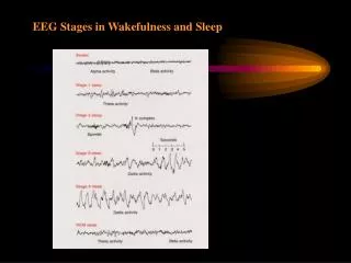
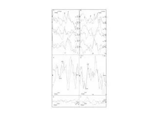
EEG activity is mostly rhytmic and of sinusoidal shape rhythm 8-13 Hz Rhythm 14-30 Hz rhythm 4-7 Hz rhythm 3 and less Hz rhythm , rolandický rytmus 8-10 Hz
Normal EEG – lokalization of graphoelement types Frontal - activity Sevření pěsti Uvolnění pěsti parietal – , rolandic rhythmus Temporal - , activity Otevření očí Zavření očí Temporo-parieto- occipital - activity Podle Faber Elektroencefalografie
Epilepsy seizure petit mal (absence) Spike and wave activity The seizure was clinically manifest as a staring spell
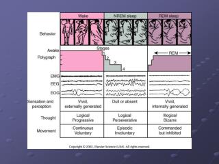
SLEEP The age-old explanation until 1940s – sleep is simply a state of reduced activity Nathaniel Kleitman in early 1950s made remarkable discovery: Sleep is not a single process, it has two distinct phases: REM sleep is characterized by Rapid Eye Movements Non-REM sleep Moruzzi in late 1950s studied reticular formation: rostral portion (above the pons) contributes to wakefulness. Neurons in the portion of RF below pons normally inhibit activity of the rostral part Sleep is an actively induced and highly organized brain state with different phases
Sleep follows a circadian rhythm about 24 hours Circadian rhythms are endogenous – persist without enviromental cues – pacemaker, internal clock – suprachiasmatic ncl. hypothalamus Under normal circumstances are modulated by external timing cues – sunlight – retinohypothalamic tract from retina to hypothalamus (independent on vision) Resetting of the pacemaker Lesion or damage of the suprachiasmatic ncl. – animal sleep in both light and dark period but the total amount of sleep is the same suprachiasmatic ncl. regulates the timing of sleep but it si not responsible for sleep itself
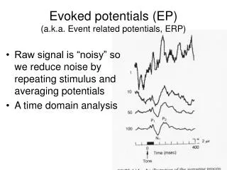
Average evoked potentials Event-related potentials Routine procedure of clinical EEG laboratories from 1980s Valuable tool for testing afferent functions EEG changes bind to sensory, motor or cognitive events
Electrical activity – electrodes placed on the patient’s scalp • Evoked electrical activity appears against a background of spontaneous electrical activity. • Evoked activity = a signal • Background activity = a noise • Signal lower amplitude than noise, it may go undetected (hidden or masked by the noise) • Solution • - by increasing amplitude of the signal – intensity of stimulation • by reducing the amount of the noise
Signal averaging Mixture of electrical activity composed of spontaneously generated voltages and the voltage evoked by stimulation Segments or epochs of equal duration Start coincides with the presentation of stimulus Duration varies from 10 to hundrets milliseconds Brain’s spontaneous electrical activity is random with respect to the signal – sum of many cycles will tend to cancel out. (to zero) The polarity of the EP will always be the same at any given point in time relative to the evoking stimulus Evoked activity will sum linearly
How to reduce the amount of the noise • Superimposition
How to reduce the amount of the noise Simplified diagram illustrating how coherent averaging enhances a low level signal (coherent = EP time locked to the evoking stimulus)
Description of waveforms: peaks (positive deflection) troughs (negative deflection) Measures: 1. Latency of peaks and troughs from the time of stimulation 2. Time elapsing between peaks and/or troughs 3. Amplitude of peaks and troughs Comparison of the patient’s recorded waveforms with normative data
Visual-evoked potentials (VEP) Anatomical basis of the VEP:
Visual-evoked potentials (VEP) Electrical activity induced in visual cortex by light stimuli Retina Rods and Cones Anatomical basis of the VEP: Bipolar neurons Ganglion cells Optic nerve Anterior visual pathways Optic chiasm Optic tract Lateral geniculate body Retrochiasmal pathways Optic radiation Occipital lobe, visual cortex
Visual-evoked potentials (VEP) Stimulus: checkerboard pattern on a TV monitor The black and white squers are made to reverse A pattern-reversal rate – from 1to 10 per second Electrodes - 3 standard EEG electrodes placed over the occipital area and a reference elektrode in a midfrontal area Analysis time (one epoch) is 250 ms Number of trials 250 , 2 tests at least to ensure that the waveforms are replicable
Normal VEP VEPs to pattern-reversal, full-field stimulation of the right eye
Abnormal VEPs Absence of a VEP Prolonged P 100 – latency - demyelination of the anterior visual pathways Amplitude attenuation - compressive lesions Prolonged P 100 only on left or right eye stimulation – lesion of the ipsilateral optic nerve Excessive interocular difference in P 100 latency – lesion of the ipsilateral optic nerve
VEPs as a tool in the diagnosis of multiple sclerosis: Excessive interocular difference in P100 latency Prolonged absolute latency Decreased amplitude Compression of optic nerve, optic chiasm (tumor of pituitary gland or optic nerve glioma) Decreased amplitude Prolonged latency of P100
Brain-stem auditory-evoked potential BAEP Short-latency somatosensory-evoked potential SSEP
Short-latency somatosensory-evoked potential SSEP Left median nerve study