Download

1 / 23
240 likes | 348 Vues
Discover in-depth insights into the causes, pathology, and treatment options for gastric cancer. Learn about key risk factors, ethiology, pathologic classifications, and available treatments to combat this condition effectively.

E N D
GASTRIC CANCER • 4% 5th position • 2nd cause of cancer related death in the world • 5 y survival rate • B:F=2:1 • More frequent in Japan, Latin America, Far East, North Europe. • Incidence is droping
Pathology • Adenocarcinoma 90% • Sarcoma • Limfoma
CAUSES • 66-75% can be prevented with diet using high quantity of fruits and vegetables and low in salty foods. • Integral cereals and green teea can reduce the incidence • Vitamin C şi carotenoidsprobably decrease the risk • Alcoholmay increase the risk of GC in cardia region • Smoking increases the risk
ETHIOLOGY • Atriphic gastritis • 9% will develop GC • Chronic inflammation – destruction of glands – lower capacity of acid secretion – intestinal metaplasia • Causes • Helicobacter pylori • Ac anti parietal cells – Biermer • Antral resection
ETHIOLOGY • Helicobacter pylori • Distal GC + association with atrophy • Appears to be protective against procimal GC • 1/ 97 infectet patients develop CG • Inf:noninf=8:1, ONLY CERTAIN FENOTYPES
ETHIOLOGY • Polyps • Hiperplastic – 80% • High risk over 0,5 cm • Adenomatos • Very high risck • Familial risk • 2-3X higher • Mutation in gene CHD1 -E-cadherina role in diferencietion and cel arhitecture • Molecular fenotype • c-met, K-sam involved in cell groth • p53 suppressor gene– 64% • cyclin E
PATHOLOGY • Intestinal • Atrophy – metaplasia - displasia – adenoma - cancer • Difuz – linitis plastica • Submucosal invasion
Macroscopic – Borrmann • Type I - polipoid well defined • Type II – polipoid with marked infiltration • Type III – ulceratio with infiltrated margins • Tip IV – linitis plastica • Microscopic – OMS • Adenocarcinoma – intestinal, difuse • Adenocarcinoma papilary • Adenocarcinoma tubular • Adenoacrcinom mucinos (>50% mucinous cells) • Signet cells carcinoma (>50% signet cells) • Adenosquamos carcinoma • Squamos cell carcinoma • Small cells carcinoma • Nondiferentiated • altele
Primary tumor (T): Tis = carcinoma in situ: intraepithelial tumor without invasion of lamina propria T1 = tumor invades lamina propria or submucosa T2 = tumor invades muscularis propria or subserosa T3* = tumor penetrates serosa (visceral peritoneum) without invasion of adjacent structures T4**,*** = tumor invades adjacent structures *A tumor may penetrate the muscularis propria with extension into the gastrocolic or gastrohepatic ligaments or into the greater or lesser omentum without perforation of the visceral peritoneum. **Structures adjacent to the stomach include the spleen, transverse colon, liver, diaphragm, pancreas, abdominal wall, adrenal gland, kidney, small intestine, and retroperitoneum. ***Intramural extension to the duodenum or esophagus is classified by the depth of greatest invasion in any of these sites, including the stomach). Regional lymph nodes (N):Include the perigastric nodes along the lesser and greater curvatures, and the nodes along the left gastric, common hepatic, splenic, and celiac arteries. N0 = no regional lymph node metastasis N1 = metastasis to 1–6 regional lymph nodes N2 = metastasis in 7–15 regional lymph nodes N3 = metastasis in more than 15 regional lymph nodes Distant metastasis (M):M0 = no distant metastasis M1 = distant metastasis
Grading • G1 - well diferentiated->95% glands • G2 – moderat diferentiated – 50-95% glands • G3 – poor diferentiated - <49% glands • Adc tubular – G1 • Adc signet cells – G3 • Adc small celss and non diferetiated – G4
CLINICA PRESENTATION • Subjectiv • General neoplastic simptoms • Dispepsia • UGI bleeding • Objectiv • Tumor palpable • Hepatomegaly, ascites, jaundice, splenomagaly • Sister Mary Joseph – sign (umbilkical nodule) • Virchow sign – left supraclavicular LN • Krukenberg –ovarian MTS • Blumer – rectal palpable mass • Trousseau – migrating flebitis • Leser-Trelat – seborheic keratitis • Lab • Anemia • Ocult bleeding • ACE, CA 19.9
CT, MRI, echoendoscopy+biopsybrush citology, laparoscopy, lapro echography
COMPLICATIONS • Bleeding • Perforation • Obstruction • Penetration
TREATMENT • SURGICAL • Rezection • R0 – complete, no microscopic tumor left • R1 – microscopic tumor left in situ • R2 – macrosocopic residual tumor • Limfadenectomy • D1 – stations 1-6 • D2 – stations 7-11 • D3 – stations 12-14 • D4 – stations 15-16 • Omentectomy
Endoscopic treatment Mucosal resection in early gastric cancer Paliative Sclerotherapy Laser destruction Stent
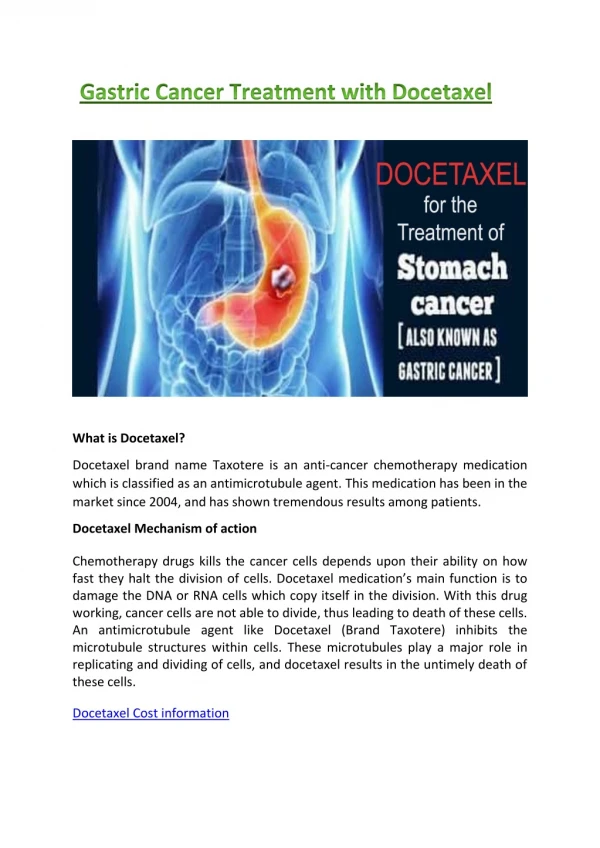
TREATMENT • Chemotherapy • Neoadjuvant / adjuvant • 5-FU, doxorubicin şi mitomycin C (FAM) • Immunochemoterapy – CHT bound to specific tumoral ATB. Ag • Radioterapy • neoadjuvant • Chemoradiation • Adjuvant • Major discussions