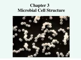
Microbial structure (2)
Structural approach : Is it a view of artifact?. Microbial structure (2). For lecture only. BC Yang. Vibrio cholerae. Staphylococcus aureus. Pseudomonas aeruginosa. Fusiform bacilli. Bacillus anthracis. Borrelia burgdorferi. Spirillum volutans. Shapes. Bacillus Coccus Coccobacillus

Microbial structure (2)
E N D
Presentation Transcript
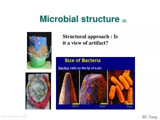
Structural approach : Is it a view of artifact? Microbial structure (2) For lecture only BC Yang
Vibrio cholerae Staphylococcus aureus Pseudomonas aeruginosa Fusiform bacilli Bacillus anthracis Borrelia burgdorferi Spirillum volutans Shapes • Bacillus • Coccus • Coccobacillus • Fusiform bacillus • Spirillum • Spirochete • Vibrio For lecture only BC Yang
Structural view of bacteria (1) • Microscopic prokaryotes (no nucleus nor membrane-bound organelles) • Contain ribosomes • Enfolding of the cell membrane carry on photosynthesis & respiration • Surrounded by protective cell wall containing peptidoglycan (protein-carbohydrate) • Many are surrounded by a sticky, protective coating of sugars called the capsule or glycocalyx For lecture only BC Yang
Structural view of bacteria (2) • One circular chromosome and some small DNA called plasmids • May have short, hairlike projections called pili on cell wall to attach to host or another bacteria when transferring genetic material • Some can move by flagella, gliding over slime they secrete ( e.g. Myxobacteria) • Some can form protective endospores around the DNA when conditions become unfavorable For lecture only BC Yang
Detail Structure For lecture only BC Yang
Structure and function For lecture only BC Yang
Differences For lecture only BC Yang
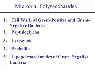
Cell envelope: Cytoplamic membrane+ cell wall+ capsules + loss slime etc…. • Gram-positive cells: • Cytoplamic membrane+ thick peptidoglycan layer + capsules • Gram-negative cells: • Cytoplamic inner membrane+ thin peptidoglycan layer + periplamic space + outer membrane + + capsules /loss slime high magnification AFM image of the surface of a single Pseudomonas putida For lecture only BC Yang
The main component of the bacterial cell wall is peptidoglycan, a hydrated, semi-rigid polymer of two sugar derivatives: N-Acteylglucosamine (G) and N-Acetylmuramic acid (M). Bound to the sugar M are amino acids: Alanine -- Glutamic acid -- Meso-diaminopimelic acid (DAP) (Gm-) or Lysine (Gm+) -- Alanine. Glucosamine? For lecture only BC Yang
Teichoic acid Are found exclusively in gram positive organisms. Are formed as polymers of glycerol or ribitol through phosphodiester linkages. http://www.cvm.uiuc.edu/courses/vp331/Structures_in_pathogenesi1.html For lecture only BC Yang
A very complex net http://www.arches.uga.edu/~kristenc/cellwall.html
Synthesis of cell wall: The assembly of the wall components begins with the synthesis of precursors in the cytoplasm, their transport across the cell membrane, and their final polymerization. Eventually, penicillin-binding proteins catalyze covalent reactions that result in the extension, cross-linking between glycan strand, morphogenessis and eventual separation of the murein sacculus. For lecture only BC Yang
透過骨架來改變形狀 BC Yang
Crescentin : creS • 430-residues protein • Distinct 7-residue repetitive pattern • coiled-coils: many fibrous proteins in eukaryotes Fluorescent image of the CreS-GFP/CreS hybrid strain BC Yang
Look into the detail For lecture only BC Yang
Gram positive • in Gm+ organisms, an interbridge exists between the Lysine molecule extending from one sugar backbone to the terminal Alanine of the second. • Gm+ organisms have cell walls that may contain as much as 90% peptidoglycan, with membrane associated proteins and Teichoic acids comprising the remaining components. For lecture only BC Yang
Gram negative • cross-linking of peptidoglycanby bond between the DAP molecule extending from one sugar backbone and the terminal Alanine of another. • typically composed of only about 10% peptidoglycan and possess an outer membrane that houses the membrane associated proteins and LPS. The peptidoglycan lies in-between the periplasm. For lecture only BC Yang
The acid-fast cell wall of Mycobacterium contains peptidoglycan, and a large amount of glycolipids such as mycolic acid, arabinogalactan-lipid comlex, and lipoarabinomannan.
Lipopolysaccharides is an amphiphile composed of three regions: O-polysaccharide (the O- or somatic-antigen), the core polysaccharide and lipid A. Lipid A is anchored in the outer membrane. LPS is also known as endotoxin. saccharide lipid Large-scale molecular dynamic simulation of a lipopolysaccharide membrane solvated in a 4.2 nm water box. For lecture only BC Yang
鱟 • Endotoxins are thermostable, lipopolysaccharide components from the cell walls of viable or nonviable gram-negative microorganisms. • Measured endotoxin will include endotoxin that was derived from: 1) Any living cells that are present, 2) Cell wall debris from dead or dying cells, and 3) Outer membrane fragments that are released during cell growth. • An endotoxin unit, EU, is equivalent to approximately 200 pico-grams of endotoxin. • The LAL (Limulus Amebocyte Lysate) test for endotoxins can be sensitive down to 0.03 EU/ml For lecture only BC Yang
Not confuse with exotoxin Tetanus toxin: (a) The x-ray crystal structure for the tetanus toxin showing how the amino acid chain is folded and (b) its calculated molecular surface showing sites 1 and 2, predicted binding sites for ligands. For lecture only BC Yang
Cytoplasmic membrane • 5-10 nm unit membrane • Absence of sterols, except mycoplasma • Semifluid state, form mesosomes • Selective permeability and transport • Electron transport /oxidative ATP generation • Bioxynthesis For lecture only BC Yang
Phospholipids • Major components of the cell membrane • Consists of two fatty acids and the third hydroxyl group of glycerol is joined to a phosphate group • being amphipathic, having both a hydrophobic (hydrocarbons tails) and a hydrophilic region (phosphate head) • In water, they self-assemble into aggregates so that the phosphate heads make contact with the water and the hydrophobic hydrocarbon tails are restricted to water-free areas (micelle or phospholipid bilayer) For lecture only BC Yang
How to get across the polar barrel Bacterial membrane proteins - OmpA (left) and GlpF (right). For lecture only BC Yang
ATP-dependent secretion systems • There are five major secretion systems. • Type I and type III secrete proteins across both the inner membrane and the cell envelope (outer membrane) in one step; secreted proteins do not make an intermediate stop in the periplasm. Type I systems are composed of far fewer components than type III systems. • Type II and type III systems share a similar cell envelope component. • The type III secretion system of Gram-negative bacterial pathogens injects virulence factors into host cells. The needle complex is drawn on the basis of its appearance in electron-microscopy images (see inset), with several ring structures that span the inner and outer membranes. Very similar to flagella system For lecture only
http://www.cdc.gov/ncidod/eid/vol2no4/mecsas.htm Bacterial secretion systems: type I, type II, and type III. For lecture only
The known type IV systems differ with respect to the route of substrate translocation. The A. tumefaciens T-DNA transfer system and the H. pylori CagA system are thought to export substrates in one step across the membrane directly to the eukaryotic cytosol. The B. pertussis Ptl system is thought to export PT in two steps across the cell envelope to the extracellular milieu. Secreted holotoxin then binds to the mammalian cell membrane. http://www.blackwell-synergy.com/links/doi/10.1046/j.1365-2958.2001.02302.x/full/ For lecture only BC Yang
Bacterial chromosome • No nuclear membrane • Small polyamines and magnesium, histone-like • Lack of mitotic apparatus; fission • ~3 x 10⁹bp (1mm long), supercoiled state, single haploid • Circular (most bacteria) or linear (Streptomyes sp.) • Associated with mesosome DNA spread of E. coli plasmid For lecture only BC Yang
Ca+2 Spores Calcium bound to dipicolinic acid Keratin-like protein coat may stay inactive several years, then re-activate when conditions favorable For lecture only BC Yang
the bacterium senses that its home or habitat is turning bad • it makes a copy of its chromosome • the rubbery cell membrane that surrounds the bacterial cell fluid begins pinching inward around this chromosome copy. • the membrane of the mother cell surrounds and swallows up the daughter cell. • between these two membranes a thick wall forms made out of stuff called peptidoglycan. • a tough outer coating made up of a bunch of proteins forms around all this, closing off the entire daughter cell. For lecture only
Capsule • Bacterial capsules outlined by India ink viewed by light microscopy. • A discrete layer of polysaccharide surrounding the cells. • Sometimes bacterial cells are embedded more randomly in a polysaccharide matrix called a slime layer or biofilm. • Polysaccharide films that may inevitably be present on the surfaces of bacterial cells, but which cannot be detected visually, are called glycocalyx. Capsule Stain of B. anthracis: Capsule appears pink, bacterial cells purple For lecture only BC Yang
Capsule • prevents desiccation of the bacteria • prevent phagocytosis by larger microorganisms and the white blood cells of invaded host organisms • help the bacteria adhere to the host substrate • assist in warding off attacks by phages For lecture only BC Yang
Pili (Fimbriae) • singular pillus • helical filaments, with ~1000 copies of the major pilin, plus one or a few copies of ~5 minor pilins. The minor proteins provide binding specificity, membrane anchoring, and adapter functions.Mu, Egelman, Bullitt, 2002. J. Bact. 284(17):4868-4874Bullitt, & Makowski, 1995. Nature 373(6510):164-167 a) Adhesionb) In pathogenesisc) In environmentd) Sex pili Sex pili also called as type IV secretion system! For lecture only BC Yang
Flagella singular flagellum A tiny motor machine Share similarity with type III secretion system For lecture only BC Yang
Swing and tumble • Each flagellum is a rigid structure • The basal body causes rotation of the structure -- like a propeller. The rate is about 12000 rpm • Energy is derived from a proton gradient. Power has been calculate to be about 10-15 amperes of current. • Average velocities are in the range of 20 - 80 µM/s. About 1/2 foot per hour. A cheetah is 4 feet long and moves about 70 mph. 25 body lengths/second. A microbe that is 2 µM long, moves 10 to 40 body lengths per second. (some bacteria are as mobile as higher animals) • Movement is a bias random walk during chemotaxis For lecture only BC Yang