EMG
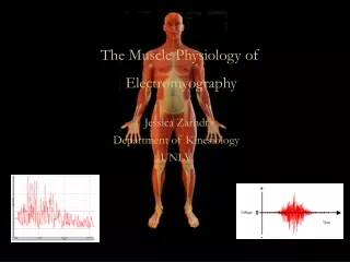
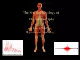
EMG. The Muscle Physiology of Electromyography. Jessica Zarndt Department of Kinesiology UNLV. EMG. Electromyography (EMG) – the measurement of electrical activity that brings about muscle contractions.

EMG
E N D
Presentation Transcript
EMG The Muscle Physiology of Electromyography Jessica Zarndt Department of Kinesiology UNLV
EMG Electromyography (EMG)– the measurement of electrical activity that brings about muscle contractions 5. Plowman SA, Smith DL. Exercise Physiology for Health Fitness and Performance. Benjamin Cummings, 2003
EMG and Muscle Physiology • Muscle Contraction • Brief Anatomical Review • Emphasis on the electrical potential • Physiological Explanation of an EMG signal • What corresponds to what we see on a signal • Physiological Factors that can Influence an EMG Signal • How do things like fiber type, size and disease affect the EMG
Series Elastic Components Tendons & Bones Fascia, Endomysium, Perimysium and the Epimysium Excitable Vs Non-Excitable Muscle tissue IS Connective is NOT Skeletal Muscle Organization
The Muscle Fiber (Cell) is excitable The Muscle Fiber is what Contracts Skeletal Muscle Organization
The Muscle Fiber at the electrophysiological level • Resting Potential – the voltage across an unstimulated cell • Muscle Cell = -90mV • Established by • Active Transport of Ions • The Na+/K+ pump • 3Na+ out / 2K+ in • Potassium Diffusion Potential • K+ diffuses in; sarcollemma is 100 x more permeable to K+ than Na+
The Muscle Fiber at the electrophysiological level • What does this mean? • High [Na+] ~140mEq/L outside the c
EMG and Muscle Physiology • How does the muscle fiber become excited & contract? • Neuro-Stimulation • Electrochemical changes in the muscle • Proteins of the muscle move-the muscle moves
1. Nervous System Signal • Originates in a Motor Neuron • Activated by conscious thought or afferent input (i.e. reflex) • Travels through the nervous system to the target muscle(s) via, depolarization (action potential) and neurotransmitters • Action Potential - a reversal in relative polarity or change in electrical potential of a cell • Neurotransmitters- chemical messengers
Action Potentialof a Neuron • Resting Potential -70mv • Excited to +35mv • The change in polarity travels down a neuron to the next • Neurotransmitter is released from terminal end
Action Potentialof a Neuron Post Synaptic Stimulation Pre Synaptic Stimulation
The Neuromuscular Junction • A specific synapse • Synapse = the junction at the terminal end of a neuron and another cell • The Neuromuscular Synapse • Motor Neuron and Muscle Cell
2. Electrochemical Changes in the Muscle 1) Ca++ are released in the terminal end of Neuron 2) Neurotransmitter is released; Acetylcholine (Ach) 3) Ach travels to receptors on muscle end plate (~50million per fiber) • Muscle End Plate – area of muscle cell innervated by neuron
Electrochemical Changes in the Muscle 4) Na+ channels open in the muscle cell -Na+ flows into the cell -Voltage begins to raise from -90mv 5) End Plate Potential- local positive potential inside a muscle fiber
5) End Plate Potential -Bidirectional -Local -Leads to AP if large enough -Usually 50-70mV Electrochemical Changes in the Muscle
Electrochemical Changes in the Muscle 6) When threshold is met in the End Plate, an Action Potential will initiate - Threshold = -55mV 7) Action Potential
EMG’s • EMG’s allow recording of the action potentials from an entire muscle (or at least significant portion of one) • The signal is a compound action potential Action potential from one skeletal muscle cell An EMG http://www.holycross.edu/departments/biology/kprestwi/phys'02/labs/emg_lab/Phys'02_L1_Intro_E-myo&FFT.pdf
EMG’S 2 1 At Rest http://www.holycross.edu/departments/biology/kprestwi/phys'02/labs/emg_lab/Phys'02_L1_Intro_E-myo&FFT.pdf
EMG’S http://www.holycross.edu/departments/biology/kprestwi/phys'02/labs/emg_lab/Phys'02_L1_Intro_E-myo&FFT.pdf
EMG’S • Evoked field potential from a single motor unit is actually (usually) triphasic • Duration is between 3 and 15 msec • Magnitude is between 20-2000 microvolts, depending on the size of the motor unit • Frequency of discharge varies from ~6 – 30 per second http://www.holycross.edu/departments/biology/kprestwi/phys'02/labs/emg_lab/Phys'02_L1_Intro_E-myo&FFT.pdf
EMG’S • Of course, when we measure an EMG, we are not recording the AP from a single motor unit, but rather we are recording from multiple cells/fibrils that: • Each generate an AP • The AP’s do not have to be in phase • Some may fire multiple times…others only once • The amplitudes of the AP’s can be different, too • The position of the electrode relative to other muscles may also cause interference The result is a signal that looks a lot like noise….but is it? http://www.holycross.edu/departments/biology/kprestwi/phys'02/labs/emg_lab/Phys'02_L1_Intro_E-myo&FFT.pdf
FOURIER ANALYSIS • We can think of an EMG as the result of the superposition of many, many waves that may or may not be in phase • By changing from the “time” domain to the “frequency” domain, we can identify the individual waves that comprise the final signal Magnitude (dB) 4 1 2 Frequency (Hz) http://www.holycross.edu/departments/biology/kprestwi/phys'02/labs/emg_lab/Phys'02_L1_Intro_E-myo&FFT.pdf
FAST FOURIER TRANSFORMS • Computational means of decomposing non-periodic signals into individual components • Fortunately, a lot programs are available to perform FFT’s (Matlab, for instance…also the Biopac software!) This FFT of an EMG shows definite peaks at 33, 45, 55, 65, 75, 90, and 94 Hz. This implies that there are large numbers of motor units firing at these frequencies! http://www.holycross.edu/departments/biology/kprestwi/phys'02/labs/emg_lab/Phys'02_L1_Intro_E-myo&FFT.pdf