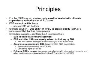
PRINCIPLES
PRINCIPLES. OF DIFFERENTIAL DIAGNOSIS AND BIOPSY TECHNIQUES. DIFFERRENTIAL DIAGNOSIS. The application of knowledge and logic to make a clinical decision Follows the hx. and phys. exam and preceeds the radiographic exam, lab studies, bx. and special tests

PRINCIPLES
E N D
Presentation Transcript
PRINCIPLES OF DIFFERENTIAL DIAGNOSIS AND BIOPSY TECHNIQUES
DIFFERRENTIAL DIAGNOSIS • The application of knowledge and logic to make a clinical decision • Follows the hx. and phys. exam and preceeds the radiographic exam, lab studies, bx. and special tests • IS BASED ON EXCLUSION and those entities that cannot be excluded make up the initial differential dx.
IMPORTANCE OF THE MEDICAL HISTORY • Pre-existing medical condition can effect surgical modalities and may be predictive of morbidity. Exam: congenital heart lesions, Htn, coagulopathies • The oral lesion may be a manifestation of systemic dz. Exam: leukemia, lichen planis, Chrohn’s dz, hairy leukoplakia • 80-90% of syst. dzs. can be discovered by thorough med hx
Notes from Previous Slide • Aplastic anemia, what process would you be concerned about? • Significant suppression of bone marrow ( a problem with WBC, RBC, and platelets) • Leukemia presents with a type of gingivitis that is very similar to ANUG • Lichen planus can be seen on the skin and other mucosal surfaces • Crohn’s diseases gives a characteristic manifestation in the mouth • Hairy leukoplakia is associated with being HIV+
LESIONS-PHYSICAL FINDINGS • Divide into 3 categories 1) Surface lesions of the mucosa and epidermis. Usually do not exceed 2mm in thickness. Divided into 3 categories based on clinical appearance: white; pigmented (brown/blue/red/black); vesicular/ulcerated/erythematous • Surface lesions – do not exceed 2mm in thickness • White lesions may be considered premalignant. • Of course when we say white we really mean leukoplakia. • Pigmented lesions can be associated with foreign body reactions (ie amalgam). These may be associated with hemorrhagic type processes. • A red lesion that is pinpoint, we may call that a petechial. • Blue lesions may be vascular, typically venous in origin. So an example of that is a varix. • Vascular lesions of course, you start to think of things like erythemamultiforme. 2) Soft tissue enlargements
LESIONS-PHYSICAL FINDINGS • 2) Soft tissue enlargements an be divided into reactive or neoplastic. Benign lesions with slow growth, not fixed, move teeth and mucosa normal. Malignant lesions may show rapid growth, ulceration, loosen teeth and systemic symptoms • Features of benign vs malignant – YOU WILL SEE THIS AGAIN • Benign • Not fixed to the underlying connective tissue (so they move) • Can move teeth and mucosa has a normal appearance • Malignant • Fixed to the underlying tissue • Ulceration is a prominent feature of a malignancy • Can loosen teeth • Usually are erosive • Show systemic signs or symptoms • A feature of a malignant process also includes paresthesia
LESIONS-PHYSICAL FINDINGS • 3)Intraosseous lesions. With the exception of lesions associated with a necrotic pulp, are less common than ST lesions. Are classified into 5 categories: cysts, odontogenic tumors, benign nonodontogenic tumors, malignant tumors, and dzs. of bone. Pain, paresthesia, growth rate, mucosa involvement are important sxs. • Intraosseous lesions are less common than soft tissue lesions • As a rule cysts are going to be fluid filled and typically will have an epithelial lining • If he were to ask: Which cyst that presents in the maxillofacial region doesn’t have an epithelial lining? • TBC (Traumatic bone cyst) • One problem with odontogenic tumors is that they can be benign of malignant. You can have benign nonodontogenic tumors, malignant tumors, and malignant diseases of bone. • Based on microscopic appearance you may have to treat a benign process like a malignant process • Example: An ameloblastoma is a benign tumor but it has many features of a malignancy and is commonly mistaken for a sarcoma? • It can mestastasize to the lungs • It has a similar lesion that appears in the brain. The ameloblastoma of the brain is a craniopharyngeoma • They are locally destructive and can cause paresthesia • Sometimes it is very difficult to decide if something is benign or malignant • The symptoms effect how you treat the lesion • Sometimes you can take a small amount of tissue, sometimes you have to take up to 1.5 cm of normal tissue to get rid of the lesion
LESIONS-CHARACTERISTICS • Duration of existence • Change in size/rate of change • Change in character vesicle/ lump/ ulcer • Sys.: pain, paresthesia,smell/ taste, adenopathy, associated constitutional changes • Any historic reason for lesion • Name a tumor that may affect smell or taste? • Salivary gland tumors, any tumors of the olfactory system, tumors of the nose and sinuses • Adenopathy implies either infection or malignancy (it is associated with these two processes) • Remember sometimes patients do things to themselves (a factitious injury)
CLINICAL EXAMINATION • Classically involves inspection, palpation, percussion, and auscultation • If you have a vascular type of sound (referred to as bruits) then that may imply that you have an AV malformation • If you have auscultate and you hear turbulence that may tell you that you have an obstruction or an AV malformation • Also evaluate: anatomic location of mass, overall physical character of lesion, color, sharpness of boundaries, consistency, presence of fluctuance/pulsation and lymph node adenopathy • If you have a mass, for example on the palate, how do you form an excisional biopsy? • The appropriate way should be parallel to the great palatine vessels rather than running across them • Biopsies in the floor of the mouth can be dangerous because of the salivary gland ducts • If you have a pulsatile feel to a mass it will almost always be an AV malformation
INDICATIONS FOR BIOPSY • Longevity of 2 weeks or longer • Persistent keratotic changes • Could be premalignant • Persistent swelling • Interference with function • Malignancy? Erythroplasia or speckled. Ulceration. Duration > 2weeks. Rapid growth rate. Bleeding with manipulation. Induration or fixation • A speckled lesion may indicate a malignancy • Induration means fixation to the underlying tissue • He’s probably going to ask if something is benign or malignant based on symptomotology
PRINCIPLES OF BIOPSY • 4 MAJOR TYPES OF BX AROUND THE ORAL CAVITY 1)Oral cytology-exfoliative cytology and oral brush cytology. • IS NOT A SUBSTITUTE FOR TRADITIONAL SCALPEL BIOPSY AND HISTOLOGY • There are some claims that you can use this to determine if something is benign or malignant, he doesn’t believe that. He believes you need actual tissue to determine that. • He hates this and can’t think of many reasons to use this other than for herpes 2)Aspiration- 18 g needle/10 cc barrel • For any osseous lucency. Also FNA (fine needle aspiration) • Can be several different things: you can stick a needle through the bone into a cyst and pull some fluid out. The color of the fluid can tell you what type of cyst you have (hemorrhagic, traumatic bone, etc) • A fine needle aspirate is a way we can take a biopsy out of the parotid gland and determine if something is benign or malignant • You can use this to decompress a cyst or to determine if something is malignant or benign
TYPES OF BIOPSY CON’T • 3) Incisional: samples a representative portion of lesion. Indicated for those lesions larger than one cm, hazardous location, suspected malignancy. Better if wedge-shaped, deep and narrow with area showing complete tissue changes. Never use necrotic tissue, gingival crevice, attached gingiva if possible • These are indicated when a mass is >1cm and when the lesion is in a very strange location (like over a salivary gland duct) and suspected malignancy • You need to do this so that if the biopsy comes back malignant the guy cutting it out knows what he is removing • These are wedge-shaped like a pie slice (deep and narrow) • For BONJ – if you send this to a pathologist they will just tell you its dead bone
TYPES OF BIOPSY CON’T • 4)Excisional: implies removal of the entire lesion so a perimeter of normal tissue surrounding the lesion(2-3mm) is included to ensure total removal. Indications include lesion < 1 cm, probable benign process. • Remove the whole mass with a perimeter of normal tissue, usually less than ½ cm • These should give you the total extent of the lesion • What happens when you have a positive margin? • You are obligated to re-excise the lesion, typically a wider local incision than what you did originally
BIOPSY-SURGICAL PRINCIPLES • Block anesthesia preferred, but if infiltration used must be ~1 cm from lesion • Apply local anesthesia around the mass, not into it • Stabilize the tissue being excised iechalazeon/suture/towel clip • When taking mucosal or lip lesions you must stabilize that lesion. • When taking something off the tongue you will use an instrument that you will screw down and it will push the lesion up cutting off the blood supply (chalazeon, can also be used on the lip) • He also places a suture on the tongue or a clip so he can pull the tongue out and hold it. That allows him to use catery if he needs to • Hemostasis: suction may not be your friend • One problem he has seen is that you take an incisional biopsy and it gets sucked up the aspirator tip. So suction can be problematic. • Incision: sharp scalpel, avoid electrocautery & most lasers. Parallel to normal course of nerves/vessels! • The incision should be sharp. He does not agree with the concept of using electrocatery to take a biopsy specimen. • You should know what the lesion is before starting to apply hot heat or light heat. • You should parallel the underlying vasculature. Never go across a nerve or vessel territory.
BIOPSY-SURGICAL PRINCIPLES • Handling of tissues: DO NOT CRUSH esp with tissue forceps/curved kelly • One problem that he sees is the specimen being crushed by the tissue forceps. This happens with a curved Kelly also. • Identify margins i.e. buccal/lingual/proximal/deep etc • When you take off a mass it is reasonable to try to identify the margins. They use sutures to do that. One suture is anterior, two are posterior, one is medial, and they will see the lateral side. • The deep is almost always noticed and typically if you have to you will put a suture on that with the label “Deep” • Specimen care: formalin/saline and not on wall of container but fully immersed • When you take a specimen what do you put it in? • Formalin is the gold standard, but you can also use saline. Saline is typically used when you have to do further studies, immunoglobulin type studies. • Primary closure is the aim • At the end you want to primarily close the wound
INTRAOSSEOUS/HARD TISSUE BIOPSY • Palpate the jaws where lesion is located-hard or spongy(implies cortical expansion) • You want to palpate the bone. If the cortical bone is soft you have cortical bone expansion. • If radiolucent-ASPIRATE-straw-colored vs air vs cheesy vsheme • If you get a cottage cheese type exudate, typically that is keratin and may be an OKC. • If you stick a needle and you get only air in your syringe it is a probable traumatic bone cyst. • If you get blood it may just mean you stuck the needle in a vessel or it may be an AV malformation. • Mucoperiosteal flaps- over sound bone and avoid major neurovascular structures • For boney lesions, when you develop your flap you want to close over bone. If your suture line is over a whole it will break down. • Removal of specimen-incisionalvsexcisional • Specimen reading will take longer • Anytime you take bone it will take a longer time to get your diagnosis back because they have to decalcify the specimen before they can read it (that takes more time). • These lesions can also be biopsied using incisionalvsexcisional processes. Why would you do that? If you have a large multilocularradiolucency, what would that be? • Can be a cyst, an odontogenic tumor (ameloblastoma or myxoma), or could be a cyst. You need to know so you need to take part of the lining. Make a flap, take some bone off the mass, aspirate it, then take a piece of tissue. You will then know what type of lesion you are dealing with when you go to remove it in total. • For an OKC you have to remove part of the bone after you take the cyst out to eradicate the daughter cyst, that is called a peripheral ostectomy. • If it is an ameloblastoma you may have to resect the jaw
WHITE SURFACE LESIONS: 1-2 Q’s • Lichen planis-bilateral distribution • A characteristic of this is that it may show bilaterality, it may show up on both sides of the midline • Nic. Stomatitis-hard palate/smoker • On the hard palate, usually have a little hard center to it, almost always associated with tobacco use • Hairy leukoplakia-immunosuppression • A sign of immunosuppression, usually associated with HIV infection. • Leukoplakias- may be premalignant • Surface debris: candidiasis, burns • Candidiasis or fungal infection, burns associated with sialicylic acid (aspirin) • Subepithelial:Fordyce granules, fibrosis • One that is common are the sebaceous glands (aka Fordyce granules), on occasion you will see fibrotic lesions that are white
LOCALIZED SURFACE PIGMENTATION • Varix, hemangioma, hematoma, ecchymosis, petechiae • Peope who may have a suspected coagulopathy may present with petechiaevsecchymosis • Ecchymosis is more a mottling of the tissue, Petechiae are pinpoint lesions • A varix of couse is a vein, you know what a hemangioma is • Melanin: Lentigo simplex, nevus, melanoma. Systemic dzsassd with melanosis incl. PeutzJegherssyndr., multiple neurofibromatosis, pregnancy, fibrous dysplasia, medication • YOU WILL SEE THIS AGAIN = patients who present with melanotic lesions of the oral mucosa, what systemic lesions may they have? • PeutzJeghers, Neurofibromatosis, Pregnancy, Fibrous dysplasia • If you have this melanosis on the skin you call it café-au-lait spots. See in all three of those (not pregnancy) • Medication – antiretrovirals tend to turn the mucosa dark, systemic antineuoplastic medications (for like colon cancer) this is a side effect, you will also see this associated with antihypertensive medications • Melanoma also presents in the mouth • The 5 year survival of melanoma in the mouth is about 0 • Amalgam tattoo
SOFT TISSUE ENLARGEMENTS • Reactive: what is tissue reacting to e.g. bacterial,physical, chemical etc • Reactive STE are usually a result of an infection or inflammation or some type of physical or chemical reaction • Neoplastic: if it appears malignant- you need to perform an incisionalbx • If its neoplastic then you need to determine if its benign or malignant • If neoplasia appears benign, divide into 4 categories 1)surface epithelial:papilloma, condyloma, keratoacanthoma,verrucous Ca. 2) mesenchymal: neurofibroma,fibroma,hemangioma, pyogenicgranuloma • Papilloma – HPV, we are starting to see a large increase in females presenting with oropharyngeal cancer, these patients can be young or old, they are running tests to see if the SCC is associated with HPV • HPV virus induced SCC patients have a better prognosis than those who get it from smoking • There will probably later be a push for males to be vaccinated • Pyogenicgranuloma – seen in pregnant patients commonly, seen in children commonly • It has a lot of features of being malignant – fast growth, fixed to the underlying tissue, can be ulcerative, bleeds all the time, but it is benign
SOFT TISSUE ENLARGEMENTS CON’T • Neoplasia con’t: 3) salivary gland neoplasia-pleomorphic adenoma 4) Cysts of soft tissues-epidermal inclusion cyst, thyroglossal duct cyst, gingival cyst, nasolabial, dermoid
SURGICAL MANAGEMENT OF CYSTS • Stepwise approach i.e. physical exam to include inspect/palpate/auscultate • Aspirate with 18g If no aspirate, lesion may be a tumor and incisional bx indicated. If air, probable traumatic bone cyst. If fluid:yellow vs opaque/cheesy vs blood
Notes on Previous Slide • You will see many cysts in your practice lifetime • Biggest problem – what type of cyst are you dealing with? • Stepwise approach ie physical exam to include inspect/palpate/auscultate • You auscultate because you are listening to see if there is bruit, a sound associated with vascular flow • Aspirate with 18g – mandated for any radiolucent lesion you approach • If no aspirate, lesion may be a solid tumor and incisional biopsy indicated • If air, probable traumatic bone cyst • Treated typically only by opening up into the cavity, ultimately this will go on to ossify once it is opened up • If fluid: yellow vs opaque/cheesy vs blood • Yellow = indicative of a dentigerous cyst • It may have a bloody exudate within it, but it will mainly be yellow • Opaque/cheese = associated with an OKC • These are important to differentiate, they have the greatest recurrence rate of all cysts • This is because OKCs have small cysts in the walls of the epithelium (daughter cysts) • The appropriate treatment for OKCs will always be removal of the cyst and then a peripheral ostectomy • That means you take about 1 mm of bone off the circumference of the cyst
CYST TREATMENT • The reason we come up with these is because if the cyst is so big at removal we put the mandible in danger of fracture, so we try to make the cyst smaller • Elimination is the aim. May be accomplished by decompression, marsupialization and enucleation • Decompression: flap, aspirate, open into cyst, maintain drainage over weeks to months. Difficult secondary to hygiene problems and if inadequate bx specimen, the lining will continue to propogate. • The aim is to make the epithelial lining of the cyst fibrotic. By doing this it will halt overall growth and then you can take out a smaller mass. • The problem with this is that the patient has to wash the area out everyday. Hygiene is a major problem • The other thing is that when you take a biopsy and every radiolucent lesion must be biopsied before providing definitive treatment. You can have a mural ameloblastoma in the lining of the cyst. Mural implies you have ingrowth within the cyst lumen. So you may not get a representative specimen. Therefore you may be decompressing an ameloblastoma, this is allowing a real tumor to propogate. • You must get a representative sample, so it may need to be a large sample. • You sew the lining of the cyst to the epithelial wall to maintain the opening
CYST TREATMENT CON’T • Marsupialization: is exteriorizing the cyst. It differs from decompression in that the roof of the cyst is removed and an synthetic tube is sewed into place and daily hygiene is maintained. The tube is prone to displacement • We now use a small plastic tube with this. It is put into the lumen of the cyst. You stick the thing in and then sew the surrounding tissue to the larger end. You can put a catheter through the tube and wash the cyst out everyday. It works well until it is dislodged. If you don’t make the flange large enough it goes into the wall and into the mandible. • Usually you will marsupialize about 4-6 months • Enucleation: Removal en toto
CYST TREATMENT CON’T • If you suspect an OKC: after enucleation, a peripheral ostectomy must be accomplished with burrs, use of chemicals (phenol or Carnoy’s solution) cryosurgery, or electrosurgery. If the OKC recurs more than 2x, resect • With an OKC you need to do a peripheral ostectomy • You can use burs (preferred method) • We tend to take a dye (methylene blue) and shoot it into the bone cavity once the cyst is out • The die penetrates about 1 mm to 1.5 mm • Other methods: • Chemicals • Carnoy’s solution – this solution has, among other things in it, formaldehyde. It doesn’t make sense to put this in living tissue. • Phenol – this is neurotoxic. If the nerve is exposed both this and Carnoy’s solution would be contraindicated. • Cryosurgery – you would take petroladium, inject it within the bone cavity and take a nitrogen probe, stick it in the middle, and turn it all the way up. The petroladium freezes and provides the peripheral ostectomy. Problem – 3 months later the patient is eating and their jaw breaks. Complication – possibility of distant fracture after performing the cryosurgery • Electrosurgery – burn the entire periphery of bone with an electrocautery
BIOPSY TECHNIQUE PRESENTATION OF CASES
Benign or malignant? • Speckled lesion – increased incidence of malignancy • Fixed • Probably the patient has a sensory deficiet • Excisional or incisional biopsy? • Incisional because the lesion is larger than 1 cm • Should be a narrow and deep biopsy, pie-shaped
Pathology?? • Malignant or benign? • Malignant • Most probable diagnosis? • Melanoma – there are satellite lesions
MALIGNANT MELANOMA • Remember: 5 year survival rate for oral melanoma is 0%
AMELOBLASTOMA-CHINA STYLE • Desmoplastic ameloblastoma • He was functioning • The tumor weighed over 2.5 lbs once taken off
Left: malignant or benign? • Malignant • Location – retromolar trigone area • Most common malignancy in the retromolar trigone area? • Squame • Top right: benign or malignant? • History of bleeding, easily traumatized, no numbness, patient is pregnant • Pyogenic granuloma – just cut it out • Bottom right: benign or malignant? • Benign, most likely gingival pathology associated with a bad tooth
Left: benign or malignant? • Benign – papilloma • What would you follow this patient for? • Cancer, what type? • Papillomas (HPV) are now associated with Oropharyngeal cancer (not oral cancer) • If they run titers on the cancer and it comes back HPV+ you have an overall better chance for survival • Top right: benign or malignant? • Probably benign • Knockle head fibroma • Excisional biopsy to remove lesion • Bottom right: benign or malignant? • Probably benign • If malignant what is the most probable etiology of the lesion? • Minor salivary gland • Lesion is a granuloma from a tongue ring
EXCISIONAL BIOPSY • Never inject into the lesion, only into the periphery • Do a pie-shaped, wedge excisional biopsy. Go all the way down to the muscle • Sew it up
There is a big vein that runs longitudinally down each side of the tongue • You should parallel vascular and neurologic structures • He doesn’t like taking biopsies from the floor of the mouth, it is right over the salivary gland ducts • Peripherative leukoplakia is hard to biopsy when in areas of the top two pictures • The bottom picture is easy to biopsy because it is in the retromolar trigone area • Don’t go too far medial, you could cut the lingual nerve
SQUAMOUS CELL CA. • Fixed lesion associated with the upper lip, most common entity would be? • Basal cell carcinoma • Fixed lesion of the lower lip, most common lesion would be? • Squamous cell carcinoma
KERATOACANTHOMA • When taking lesions off on the face, we have to put the incision in areas where we get a natural skin crease • Here he put it in the nasolabial fold • He made a curvilinear incision here • You do not want to violate the vermillion border when doing biopsies
PLEOMORPHIC ADENOMA • Concepts to remember when dealing with the palate • Vasculature – the greater palatine artery • The nerve • If you violate the hard and soft palate what do you need to worry about? • The constrictor muscles, the patient will have a problem with swallowing • If you are doing a large palatal excisional biopsy, you almost always have to have a palatal stent or obturator for patient comfort and to eat
PATIENT COMFORT DURING HEALING PHASE IMPORTANT • When you take off a pleomorphic adenoma you need a cuff of surrounding tissue • Benign – 0.5-1cm normal margin • Malignancy – 1-1.5cm margin • You can use some intrawound packing now. There are some good hemostatic agents • When it heals it almost always heals with a scar across your vibrating line, this would make it difficult to wear a denture
CYST THERAPY • Fluid appearance from a cyst • Yellow-reddish exudate when aspirating 18 GAUGE ASPIRATION
CENTRAL GIANT CELL GRANULOMA • Got blood out on aspiration • Got 25cc and then the blood stopped • There is a large mass there, you can see the mental nerve on the bottom right
The mass was taken completely out, it was a pretty good sized tumor • Once taken out, if it is found to be benign you can think about doing an immediate reconstruction
Canalicular Adenoma • A lesion that in particular is only associated with the upper lip