
Comprehensive Guide to Adrenal Incidentaloma Management
E N D
Presentation Transcript
Definition • An adrenal incidentaloma is an adrenal mass detected on imaging not performed for suspected adrenal disease. • The imaging study is not done for symptoms related to adrenal hormone excess (e.g. pheochromocytoma, Cushing’s or Conn’s syndrome) or an otherwise suspected adrenal mass . • Screening imaging in patients with a hereditary syndrome leading to adrenal tumors is outside the definition of an adrenal incidentaloma. • Adrenal masses discovered on an imaging study performed during tumor evaluation for extra-adrenal malignancies (‘tumor staging’ or follow-up) do not meet the strict definition of adrenal incidentaloma.
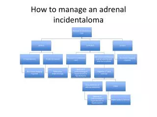
Assessment of the risk of malignancy • We recommend aiming to establish if an adrenal mass is benign or malignant at the time of initial detection. • We recommend that all adrenal incidentalomasundergo an imaging procedure to determine if the mass is homogeneous and lipid-rich and therefore benign (⊕OOO). For this purpose, we primarily recommend the use of noncontrastCT (⊕OOO).
We suggest that if the noncontrast CT is consistent with a benign adrenal mass (Hounsfield units ≤10) that is homogeneous and smaller than 4 cm, no further imaging is required (⊕OOO).
If the adrenal mass is indeterminate on noncontrast CT and the results of the hormonal work-up do not indicate significant hormone excess, there are three options that should be considered by a multidisciplinary team acknowledging the patient’s clinical context: • immediate additional imaging with another modality, • interval imaging in 6–12 months (noncontrast CT or MRI) • or surgery without further delay.
We recommend against the use of an adrenal biopsy in the diagnostic work-up of patients with adrenal masses unless there is a history of extra-adrenal malignancy .
Assessment for hormone excess • We recommend that every patient with an adrenal incidentaloma should undergo careful assessment including clinical examination for symptoms and signs of adrenal hormone excess. • We recommend excluding pheochromocytoma by measurement of plasma-free metanephrinesor urinary fractionated metanephrines.
In patients with concomitant hypertension or unexplained hypokalemia, we recommend the use of the aldosterone/renin ratio to exclude primary aldosteronism. • We suggest measurement of sex hormones and steriodprecursors in patients with imaging or clinical features suggestive of adrenocortical carcinoma.
Follow-up of patients not undergoing adrenalsurgery after initial assessment • We suggest against further imaging during follow-up in patients with an adrenal mass <4 cm with clear benign features on imaging studies (⊕OOO). • Some panel members argued that one follow-up imaging (noncontrast CT or MRI) after 6–12 months might be considered in lesions >4 cm.
In patients with an indeterminate adrenal mass (by imaging), opting not to undergo adrenalectomyfollowing initial assessment, we suggest a repeat noncontrast CT or MRI after 6–12 months to exclude significant growth (⊕OOO). • We suggest surgical resection if the lesion enlarges by more than 20% (in addition to at least a 5 mm increase in maximum diameter) during this period. • If there is growth of the lesion below this threshold, additional imaging again after 6–12 months might be performed.
We suggest against repeated hormonal work-up in patients with a normal hormonal work-up at initial evaluation unless new clinical signs of endocrine activity appear or there is worsening of comorbidities (e.g. hypertension and type 2 diabetes) (⊕OOO).
Small Phaeo/sPGL in their early stages of development are generally associated with few or mild symptoms. • Also large Phaeo/sPGL with extensive internal necrotic/haemorrhagicareas may be associated with a mild clinical picture.
Patients with adrenaline-secreting tumors present more often with paroxysmal signs and symptoms, but outside the secretory crisis the patient can present with normotension and clinical silence. • In patients with predominantly noradrenaline-secreting tumorsthe clinical picture can be mild, resembling essential hypertension. Insome of them , the continuously higher plasma concentrations of noradrenaline may induce a down-regulation of adrenoceptors, which cause amilder clinical picture, sometimes to a normal blood pressure profile.
Rarely, large tumours may be asymptomatic even in the absence of intra-tumoural necrosis. Such tumours, which have been reported in patients with succinate dehydrogenase subunit B (SDHB) mutations , have an undifferentiated catecholamine biosynthetic phenotype, contain low or negligible concentrations of catecholamines and can reach large sizes before secreting sufficient amounts of catecholamines to produce signs and symptoms.
PGLs that do not produce or secrete catecholamines : • Parasympathetic in origin and located in the head and neck region (HNPGL) or in the upper/anterior mediastinum. • Phaeo/PGL that do not secrete catecholamines but metabolize them to inactive compounds. • Do not even synthesize or contain catecholamines as a result of a defect in tyrosinehydroxylase.
Normotensive Incidentally DiscoveredPheochromocytomas Display Specific Biochemical,Cellular, and Molecular Characteristics