Download
1 / 79
940 likes | 2.22k Vues
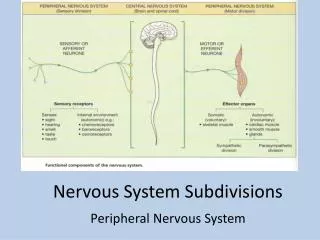
Subdivisions of Nervous System. Functions of the Nervous System. Sensory input – gathering information To monitor changes occurring inside and outside the body Changes = stimuli Integration To process and interpret sensory input and decide if action is needed Effect or Action

E N D
Functions of the Nervous System • Sensory input – gathering information • To monitor changes occurring inside and outside the body • Changes = stimuli • Integration • To process and interpret sensory input and decide if action is needed Effect or Action The result of the integration. A reflex or response.
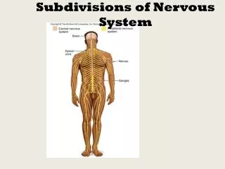
Subdivisions of Nervous System Two major anatomical subdivisions • Central nervous system (CNS) • brain and spinal cord enclosed in bony coverings • Peripheral nervous system (PNS) • Every Nerve outside of the CNS! • NERVE: a bundle of neurons • GANGLIAN: a cluster of neuron bodies ( resembles a knot)
Where Are My Nerves? • We have : • 12 Cranial Nerves • 33 Spinal Nerves
Functions of The Nervous System • Regulates Internal Body Metabolism (Body temp., urine volume, blood volume, gas exchange, circulation movement) • Links To External Environment ( Interpreter) Sensory Devices-seeing, smelling, hearing (pressure, hot cold) Emotion Response to external environment ( these drives satisfy physical needs to preserve homeostasis) thirst, hunger,rest sexuality
Overview of Nervous System • Endocrine and nervous system maintain internal coordination • endocrine = chemical messengers (hormones) delivered to the bloodstream • nervous = three basic steps • sense organs receive information • brain and spinal cord determine responses • brain and spinal cord issue commands to glands and muscles
Functional Divisions of PNS • Sensory (afferent) divisions (receptors to CNS) (Leads to…with stimuli) • visceral sensory and somatic sensory division • Motor (efferent) division (CNS to effectors) • visceral motor division (ANS) effectors: cardiac, smooth muscle, glands • sympathetic division (action) • parasympathetic division (digestion) • somatic motor division effectors: skeletal muscle
Cells of the Nervous System Are Specialized and Are of Several Types
Neuroglial Cells of CNS THESE ARE SUPPORT CELLS THAT OUTNUMBER THE NEURONS
Astrocyte • Astrocytes • Abundant, star-shaped cells • Brace neurons w/ their perivascular feet. • Form barrier between capillaries and neurons • Control the chemical environment of the brain; Secrete Neural Growth factors • Convert Glucose to lactate
ASTROCYTES ALSO……. Most common glial cell in CNS Form blood-brain barrier Help with ion uptake Help with neurotransmitter uptake Many glucose transport carriers, which help move glucose from blood to brain
Neuroglia: Support Cells • Ependymal cells • Lines the ventricles of the brain Spaces filled with CSF)and spinal Chord • Produced Cerebral Spinal Fluid • Assists in the Circulation of CSF Figure 7.3b–c
Microglia • Phagocytic cells that engulf and destroy microbes and debris in the CNS. • Active in response to immune response or injury • Spider-like in appearance
Neuroglia: Support Cells • Satellite cells • Protect sensory neuron cell bodies in the dorsal root ganglia • Schwann cells • Form myelin sheath in the peripheral nervous system (PNS) Figure 7.3e
Oligodendrocyte :forms myelin in the CNS • Myelin: composed from lipids (contains cholesterol) • Creates nodes of ranvier • This insulation causes saltatory conduction, a more rapid conduction than w/out insulation
Schwann Cell or Neurolemmocyte • The neurolemmocyte is the neuroglia cell that surrounds the axon of the neuron in the PNS. Here
Schwann Cell or Neurolemmocyte • IN FACT THIS CELL produces the myelin sheath around the axon in the PNS…yeah!! • Neurolemmna- The outer nucleated layer cytoplasmic layer of the neurolemmocyte • Nodes of Ranviar- Periodic Gaps between the myelin sheath.
The Structural Cell of the Nervous System: The Neuron SPECIAL CHARACTERISTICS: • loses its ability to divide (no centrioles) There are exceptions to this rule…hippocampus and olfactory neurons. • consumes a great deal of energy; many mitrochondria are present • THEY CAN BE VERY LARGE! Some are as long as one meter.
Which Are Found in CNS? • Ependymal • Microglia • Astrocytes • Oligodendrocytes • Satellite and Schwann Cells are found in PNS
The Neuron : Structural Cell of NS • The Basic information processing unit of the NS. • Conducts impulses from one part of body to another • Has 3 distinct parts: • Cell Body ( Soma) • Dendrites • Axon
Structure of a Neuron • Cell body = perikaryon = soma • single, central nucleus with large nucleolus • cytoskeleton of microtubules and neurofibrils (bundles of actin filaments) • compartmentalizes RER into Nissl bodies Vast number of short dendrites • for receiving signals • Single axon (nerve fiber) arising from axon hillock for rapid conduction • axoplasm and axolemma and synaptic vesicles
Variation in Neural Structure • Multipolar neuron • most common • many dendrites/one axon • Bipolar neuron • one dendrite/one axon • olfactory, retina, ear • Unipolar neuron • sensory from skin and organs to spinal cord • TOP 3 Use # of projections from soma to classify: Know where they are found. • Anaxonic neuron • many dendrites/no axon • help in visual processes
Fundamental Types of Neurons • Sensory (afferent) neurons • detect changes in body and external environment • information transmitted into brain or spinal cord • Interneurons (association neurons) • lie between sensory and motor pathways in CNS • 90% of our neurons are interneurons • process, store and retrieve information • Motor (efferent) neuron • send signals out to muscles and gland cells • organs that carry out responses called effectors
It All Starts at The Sensory Receptor • Sensory neurons- or Afferent neurons collect information from the external environment. • Bring information to the CNS
Afferent Neuron – Moving away from a central organ or pointRelays messages from receptors to the CNS
Sensory neurons Carries impulses from receptors e.g pain receptors in skin to the CNS( brain or spinal cord)
Motor neuron Carries impulses from CNS to effector e.g. muscle to bring about movement or gland to bring about secretion of hormone e.g ADH
Parts of a neuron Cell body- contains the nucleus and other organelles Dendrites- transmit electrical impulses TO the cell body Axon- transmits impulse AWAY from the cell body axons can be several feet long “Axonal hillock” is located near the cell body nerve impulses originate there
Axon- A single long thin extension that sends impulses to another neuron or tissue Axon Hillock- Where the axon originates ( no nissil bodies. Axon Collaterals- side branches along the length of the axon Axon Terminals- many tiny end filaments derived from branching of an axon and collaterals. Synaptic Bulb-bulb-like structure at the end of axon terminal ; contains vesicles that store neurotransmitters Axoplasm- the cytoplasm found in the neuron Axolemma- neuron’s plasma membrane
Properties of Neurons • Excitability (irritability) • ability to respond to changes in the body and external environment called stimuli • Conductivity • produce traveling electrical signals • Secretion • when electrical signal reaches end of nerve fiber, a chemical neurotransmitter is secreted
Presynaptic To Postsynaptic Membrane Communication • Neurotransmitters are chemicals released into the synaptic cleft at the synapse (region of contact) • Stored in vesicles, NTs, when released, activate receptors at postsynaptic membrane. • Acetylcholine, serotonin, GABA, Dopamine (examples of NTs)
Synapses between Neurons • First neuron releases neurotransmitter onto second neuron that responds to it • 1st neuron is presynaptic neuron • 2nd neuron is postsynaptic neuron • Synapse may be axodendritic,axosomaticor axoaxonic ( What do you think This Means?) • Number of synapses on postsynaptic cell variable • 8000 on spinal motor neuron • 100,000 on neuron in cerebellum
How Do Neurons Work? How Do they React, Conduct, Secrete? UNDERSTANDING THE NERVE IMPULSE
Nerve Impulse Relies on the ELECTROCHEMISTRY of the Plasma Membrane
Before Neurons Conduct or “Propagate” an impulse they are in the RESTING state • This means no impulse is being fired. • In order for a impulse to be generated, a stimulus must be provided. This will cause the ions Na+, via channels, to pass from outside of membrane to inside. • If THRESHOLD is met a NERVE IMPULSE will carry.
SENDING INFORMATION: NERVE IMPULSE Sending Information- The nerve impulse refers to the series of separate action potentials that take place segment by segment as they move down the length of the axon. All-or-None law – If an action potential starts at the beginning of the Axon, the action potential will continue at the same speed segment to segment to the very end of the axon.
Nerve Action • A. Resting potential – a non-firing neuron in which the inside is more negative than the outside of the neuron
Gates • special passageways for these two ions that are commonly referred to as GATES , • there are SODIUM GATES and POTASSIUM GATES .These gates represent the only way that these ions can pass through the nerve cell membrane. IN A RESTING NERVE CELL MEMBRANE, all the sodium gates are closed and some of the potassium gates are open. AS A RESULT, sodium cannot diffuse through the membrane & largely remains outside the membrane. HOWEVER, some potassium ions are able to diffuse out.
Two ions are responsible: sodium (Na+) and potassium (K+). • An unequal distribution of these two ions occurs on the two sides of a nerve cell membrane because carriers actively transport these two ions: sodium from the inside to the outside and potassium from the outside to the inside. • AS A RESULT of this active transport mechanism (commonly referred to as the SODIUM - POTASSIUM PUMP, there is a higher concentration of sodium on the outside than the inside and a higher concentration of potassium on the inside than the outside.
Resting Membrane Potential. Its Tested ….Like the Battery. • With one electrode placed inside a neuron and the other outside, the voltmeter is 'measuring' the difference in the distribution of ions on the inside versus the outside. And, in this example, the voltmeter reads -70 mV (mV = millivolts). In other words, the inside of the neuron is slightly negative relative to the outside. This difference is referred to as the Resting Membrane Potential. How is this potential established?
REMEMBER Which side is positive and which is negative • OUTSIDE: POSITIVE ( ABUNDANCE OF SODIUM IONS DO THIS. • INSIDE: NEGATIVE We derive at our NET negative resting figure from the INSIDE