Download

1 / 18
180 likes | 373 Vues
Vivek Murthy Intern Boot Camp – 2013. Shortness of breath. Overview. Dyspnea Mechanisms (really briefly) Questions to ask Evaluating the patient Differential diagnosis Workup Treatment options Considering a higher level of care. Definition. Dyspnea (symptom)

E N D
Vivek Murthy Intern Boot Camp – 2013 Shortness of breath
Overview • Dyspnea • Mechanisms (really briefly) • Questions to ask • Evaluating the patient • Differential diagnosis • Workup • Treatment options • Considering a higher level of care
Definition • Dyspnea (symptom) • per the American Thoracic Society: “…subjective experience of breathing discomfort that consists of qualitatively distinct sensations that vary in intensity. The experience derives from interactions among multiple physiological, psychological, social and environmental factors and may induce secondary physiological and behavioral responses.”
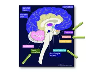
Mechanisms of dyspnea • Sensory afferent • Chemoreceptor (carotid body, medulla) - hypoxemia, hypercapnia, acidemia • Mechanoreceptor (lungs) – bronchospasm • Juxtacapillary “J” receptors – elevated PA pressures, pulmonary edema • Metaboreceptors (skeletal muscles) • Motor efferent (ventilatory pump), feedback to sensory limb • Decreased lung compliance • Respiratory muscle weakness • Anxiety • Either altered perception of physiologic data or • Tachypnea in the setting of airflow limitation (e.g. asthma) causing hyperinflation
Causes of hypoxia • With an increased A-a gradient • RL Shunt (PFO, pulmonary AVM, severe atelectasis, etc.) • V/Q mismatch (PE, alveolar process, to some extent present in each lung zone) • Diffusion limitation (ILD) • Without an A-a gradient • Hypoventilation: neuromuscular disease, AMS (narcotics), OSA, Pickwickian breathing • Low inspired FiO2 (even on Tower 11, this will not be an issue in the Cleve)
“This is Orange team returning a page” You are intern NF. A nurse calls reporting that a patient is short of breath. Questions? • Heart rate • tachycardia (arrhythmia, ST 2/2 edema or PE, SIRS/sepsis) • Blood pressure • Severe HTN (flash pulmonary edema), hypotension (large PE, MI, sepsis) • Temperature • Fever: PNA, VTE • Patient’s appearance/mental status/new complaints (eg emesis, CP) • Hypercapnia, hypoventilation, aspiration event • Current/baseline Oxygen requirement • Is pt supposed to be on 4L at baseline and currently on RA? Supposed to be on BiPap but not? Simple fix! • Recent meds/transfusions/IV fluids • Consider narcoticshypoventilation, TRALI, continuous IV fluids w/pulmonary edema Always evaluate the patient yourself! Be sure to document the scenario in a brief note.
En route to the patient • Things to determine as you walk briskly, but confidently to the patient’s room: • Who is this and why are they here? • Relevant diagnoses • Recent procedures (surgeries, thoracic/endovascular procedures, EP procedures?) • Risk factors for VTE? On DVT prophylaxis? • Oxygen requirement, baseline SaO2 • Medications: sedating meds? Respiratory meds? Due for diuretics? • I/O in the last 24-48h • Labs: Bicarb? Hemoglobin trend? Prior ABGs? BNP? • Prior imaging • Code Status, Goals consistent with elective intubation or ICU transfer?
At the bedside • Sick or not Sick? Your initial impression is very important. • Is the patient lying comfortably in bed? Supine vs tripod? Accessory muscle use? • Speaking normally or conversationally dyspneic? • What is the patient’s mental status? • Questions for the patient: • When did this start? • Have you had this before? • Cough? • Chest pain? • Positional (e.g. orthopnea? platypnea?) • Persistent vs intermittent? • Dysphagia, emesis, coughing after recent meal?
At the bedside • Vitals • Focused physical exam: • JVP • Lung exam: Stridor? Rales? Wheeze? Diminished air entry—unilateral/bilateral? • Cardiac exam: rate/rhythm? S3? Loud S2? MR? • Abdominal exam: markedly distended? • LE edema: unilateral/bilateral, signs for DVT?
Causes of acute dyspnea to consider • Pulmonary edema • PE • COPD/Asthma exacerbation • Pneumonia, especially with mucous plugging • Pneumothorax • Aspiration (initially pneumonitis, may progress to PNA) • ARDS • Sepsis, significant metabolic acidosis • MI • Pericardial effusion • Neuromuscular disease (severe, e.g. myasthenic crisis) • Anemia
Causes of pulmonary edema to consider • Cardiogenic pulmonary edema • LV failure ± acute MI • Acute valve failure (especially MR) • Tachyarrhythmia • Non-cardiogenic pulmonary edema • Hypertensive emergency • Aspiration, inhalation • Re-expansion pulmonary edema • ARDS • IL-2 infusion
Initial diagnostic tests • CXR • Diffuse process (alveolar vs interstitial) • Focal infiltrate (PNA, atelectasis, aspirate, infarction) • Extrapulmonary findings (pleural/pericardial effusion, PTX) • EKG • Ischemic changes • Arrhythmias • Signs of Right heart strain • ABG • More on this in the next slide • Well’s Criteria: consider D-dimer vs CT Angiography vs V/Q scan • Consider CBC, RFP, BNP, Troponin
Initial diagnostic tests • ABG • Respiratory acidosis or alkalosis Acute vs chronic? Acute on chronic? • For every change in pCO2 of 10 (deviating from norm of 40) • pH changes 0.08 in the acute setting • pH changes 0.04 in chronic CO2 retention • Metabolic acidosis or alkalosis Respiratory compensation? Appropriate or inappropriate? • Winter’s formula: measured Bicarb x 1.5 +8 ± 2 = expected pCO2 • If pCO2 is lower, there is an independent respiratory alkalosis • If pCO2 is higher, there is also respiratory acidosis • Does the pCO2 make sense given the pt’s degree of tachypnea? • If the RR is 40 and the pCO2 is normal, you should be concerned that the patient is tiring. • If you cannot obtain an ABG, a VBG is acceptable to check pCO2 and pH.
“So…what do you want to do, doc?” • Lasix (once, q2, or gtt) • Bronchodilators • Antibiotics (see Dr. Doucet’s talk for details) • Afterload reduction • Treating an MI (to be discussed by Dr. Tam) • Heparin gtt • Oxygen (nasal cannula, VM, NRB, NIPPV) • Suctioning (Nasotracheal suctioning for mucous plugs) • Anxiolytics? • ICU transfer
Oxygen therapy • Nasal cannula: 24-44% FiO2 • Each “liter” is ~4% above 20% (1L is 24%, 2L 28%, 3L 32%, 4L 36%, 5L 40%) • Venturi mask: ~50% • Non-rebreather: 100% • AmbuBag (Bag Valve Mask): 100% with manual ventilator support • Continuous positive airway pressure (CPAP): useful in hypoxia • Reduces pulmonary edema (afterload reduction, direct effect on hydrostatic pressure) • Bi-level positive airway pressure (BiPap): useful in hypercapnia • Gradient between iPap/ePaphelps offload CO2 • Endotracheal intubation • If patient is unable to protect their airway, vomiting (can’t use NIPPV), or…you think they need it.
Rally your forces! • There are always at least 3 senior residents in the hospital—do not hesitate to call for help • Sick or Not Sick—if “Sick”, consider calling a Code White or M&R • You will have support from an experienced MICU RN • 1:1 nursing until the acute issue is addressed • Facilitates ICU transfer • At UH, there is a Pulmonary fellow in-house at all times • ICU transfer for new NIPPV, patients who may require intubation, or closer monitoring