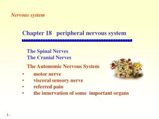
Overview of Spinal Nerves and Plexuses
560 likes | 602 Vues
Explore the thirty-one pairs of spinal nerves connected to the spinal cord, including structure, ramus branches, plexuses, and dermatomes/myotomes. Discover the composition and functions of the twelve pairs of cranial nerves in the brainstem.

Overview of Spinal Nerves and Plexuses
E N D
Presentation Transcript
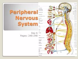
SPINAL NERVES Overview Thirty-one pairs of spinal nerves are connected to the spinal cord (Figure 14-1) No special names; numbered by level of vertebral column at which they emerge from the spinal cavity Eight cervical nerve pairs (C1 through C8) 12 thoracic nerve pairs (T1 through T12) Five lumbar nerve pairs (L1 through L5) Five sacral nerve pairs (S1 through S5) One coccygeal nerve pair
SPINAL NERVES (cont.) Lumbar, sacral, and coccygeal nerve roots descend from point of origin to the lower end of the spinal cord (level of first lumbar vertebra) before reaching the intervertebral foramina of the respective vertebrae, through which the nerves emerge Cauda equina describes the appearance of the lower end of the spinal cord and its spinal nerves as a horse’s tail
SPINAL NERVES (cont.) Structure of spinal nerves Each spinal nerve attaches to spinal cord by a ventral (anterior) root and a dorsal (posterior) root Dorsal root ganglion: swelling in the dorsal root of each spinal nerve All spinal nerves are mixed nerves
SPINAL NERVES (cont.) Ramus One of several large branches formed after each spinal nerve emerges from the spinal cavity (Figure 14-2) Dorsal ramus supplies somatic motor and sensory fibers to smaller nerves that innervate the muscles and skin of the posterior surface of the head, neck, and trunk
SPINAL NERVES (cont.) • Ventral ramus • Structure is more complex than that of dorsal ramus • Autonomic motor fibers split from the ventral ramus and head toward a ganglion of the sympathetic chain • Some autonomic fibers synapse with neurons that continue on to autonomic effectors through splanchnic nerves; others synapse with neurons whose fibers rejoin the ventral ramus • Sympathetic rami: splitting and rejoining of autonomic fibers • Motor and sensory fibers innervate muscles and glands in the extremities and lateral and ventral portions of neck and trunk
SPINAL NERVES: PLEXUSES (cont.) Nerve plexuses Plexus: complex network formed by the ventral rami of most spinal nerves (not T2 through T12), subdividing and then joining together to form individual nerves Fibers from several different rami join together to form individual nerves
SPINAL NERVES: PLEXUSES (cont.) Four major pairs of plexuses Cervical plexus (Figure 14-3) Located deep within the neck Composed of ventral rami of C1 through C4 and a branch of the ventral ramus of C5 Individual nerves emerging from cervical plexus innervate the muscles and skin of the neck, upper shoulders, and part of the head Phrenic nerve exits the cervical plexus and innervates the diaphragm Brachial plexus (Figure 14-4) Located deep within the shoulder Composed of ventral rami of C5 through T1 Individual nerves emerging from brachial plexus innervate the lower part of the shoulder and the entire arm
SPINAL NERVES: PLEXUSES (cont.) Lumbar plexus (Figure 14-5) Located in the lumbar region of the back in the psoas muscle Formed by intermingling fibers of L1 through L4 Femoral nerve exits the lumbar plexus, divides into many branches, and supplies the thigh and leg Sacral plexus and coccygeal plexus (Figure 14-5) Located in the pelvic cavity in the anterior surface of the piriformis muscle Formed by intermingling of fibers from L4 through S4 Tibial, common peroneal, and sciatic nerves exit the sacral plexus and supply nearly all the skin of the leg, posterior thigh muscles, and leg and foot muscles Coccygeal plexus arises from S5 and S4 and supplies the skin over the coccyx bone
SPINAL NERVES (cont.) Dermatomes and myotomes (Figure 14-6) Dermatome: region of skin surface area supplied by afferent (sensory) fibers of a given spinal nerve (Figure 14-7) Myotome: skeletal muscle or muscles supplied by efferent (motor) fibers of a given spinal nerve (Figure 14-8)
CRANIAL NERVES Overview (Tables 14-2 and 14-3) 12 pairs of cranial nerves connect to the brain, mostly on the brainstem (Figure 14-9) Identified by name (determined by either distribution or function) or number (order in which they emerge, anterior to posterior) or both Composed of bundles of axons Mixed cranial nerve: axons of sensory and motor neurons Sensory cranial nerve: axons of sensory neurons only Motor cranial nerve: mainly axons of motor neurons and a small number of sensory fibers (proprioceptors)
CRANIAL NERVES (cont.) Olfactory nerve (I) Composed of axons of neurons whose dendrites and cell bodies lie in nasal mucosa and terminate in olfactory bulbs Carries information about sense of smell Optic nerve (II) Composed of axons from the innermost layer of sensory neurons of the retina Carries visual information from the eyes to the brain
CRANIAL NERVES (cont.) Oculomotor nerve (III) Fibers originate from cells in the oculomotor nucleus and extend to some of the external eye muscles Efferent autonomic fibers are also present, extending to the intrinsic muscles of the eye to regulate amount of light entering eye and help focus on near objects
CRANIAL NERVES (cont.) • Trochlear nerve (IV) • Motor fibers originate in cells of the midbrain and extend to the superior oblique muscles of the eye • Means “pulley” since the muscle passes through a pulley like ligament
CRANIAL NERVES (cont.) Trigeminal nerve (V) (Figure 14-10) Has three branches: ophthalmic nerve, maxillary nerve, and mandibular nerve Sensory neurons carry afferent impulses from skin and mucosa of head and teeth to cell bodies in the trigeminal ganglion Motor fibers originate in trifacial motor nucleus and extend to the muscles of mastication through the mandibular nerve
CRANIAL NERVES (cont.) • Abducens nerve (VI) • Motor nerve with fibers originating from a nucleus in the pons on the floor of the fourth ventricle and extending to the lateral rectus muscles of the eye • Abducts the eye
CRANIAL NERVES (cont.) Facial nerve (VII) Motor fibers originate from a nucleus in lower part of pons and extend to superficial muscles of the face and scalp (Figure 14-11) Autonomic fibers extend to submaxillary and sublingual salivary glands Also contains sensory fibers from taste buds of anterior two thirds of the tongue
CRANIAL NERVES (cont.) Vestibulocochlear nerve (VIII) Two distinct divisions that are both sensory: vestibular nerve and cochlear nerve Vestibular nerve fibers originate in the semicircular canals in inner ear and transmit impulses that result in sensations of equilibrium Cochlear nerve fibers originate in organ of Corti in the cochlea of the inner ear and transmit impulses resulting in sensations of hearing
CRANIAL NERVES (cont.) Glossopharyngeal nerve (IX) Composed of sensory, motor, and autonomic nerve fibers Supplies fibers to tongue, pharynx, and carotid sinus (Figure 14-12) Vagus nerve (X) Composed of sensory and motor fibers with many widely distributed branches Sensory fibers supply pharynx, larynx, trachea, heart, carotid body, lungs, bronchi, esophagus, stomach, small intestine, and gallbladder (Figure 14-13) Somatic motor fibers innervate the pharynx and larynx and are mostly autonomic fibers
CRANIAL NERVES (cont.) Accessory nerve (XI) Motor nerve that is an “accessory” to the vagus nerve Innervates thoracic and abdominal viscera, pharynx, larynx, trapezius, and sternocleidomastoid (Figure 14-14) Hypoglossal nerve (XII) Motor fibers innervate the muscles of the tongue Contains sensory fibers from proprioceptors in muscles of the tongue
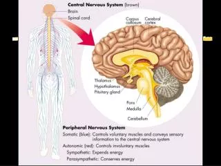
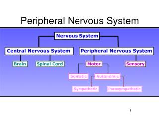
DIVISIONS OF THE PERIPHERAL NERVOUS SYSTEM Two functional divisions of the peripheral nervous system Afferent (sensory) division Efferent (motor) division Efferent division is divided further into the somatic motor nervous system and the efferent portions of the autonomic nervous system
DIVISIONS OF THE PERIPHERAL NERVOUS SYSTEM (cont.) Basic principles of somatic motor pathways Somatic nervous system includes all voluntary motor pathways outside the central nervous system Somatic effectors: skeletal muscles
DIVISIONS OF THE PERIPHERAL NERVOUS SYSTEM (cont.) Somatic reflexes Nature of a reflex Reflex: action that results from a nerve impulse passing over a reflex arc; predictable response to a stimulus Cranial reflex: center of reflex arc is in the brain Spinal reflex: center of reflex arc is in the spinal cord Reflex consists of either muscle contraction or glandular secretion Somatic reflex: contraction of skeletal muscles Autonomic (visceral) reflex: either contraction of smooth or cardiac muscle or secretion by glands
DIVISIONS OF THE PERIPHERAL NERVOUS SYSTEM (cont.) Somatic reflexes of clinical importance: reflexes deviate from normal in certain diseases, and reflex testing is a valuable diagnostic aid (Figure 14-16) Knee jerk (also known as patellar reflex): extension of the lower leg in response to tapping the patellar tendon; tendon and muscles are stretched, stimulating muscle spindles and initiating conduction over a two-neuron reflex arc (Figure 14-15) Ankle jerk (also known as Achilles reflex): extension of the foot in response to tapping the Achilles tendon
Plantar reflex: curling of all toes and a slight turning in and flexion of the anterior part of the foot in response to stimulation of the outer edge of the sole; compare to Babinski sign Babinski sign: extension of great toe, with or without fanning of other toes, in response to stimulation of outer margin of sole of foot; present in normal infants until approximately 1½ years of age and then becomes suppressed when corticospinal fibers become fully myelinated; in human beings older than 1½ years, a positive Babinski reflex is one of the pyramidal signs indicating destruction of corticospinal (pyramidal tract) fibers DIVISIONS OF THE PERIPHERAL NERVOUS SYSTEM: SOMATIC REFLEXES (cont.)
Corneal reflex: winking in response to the cornea being touched; mediated by reflex arcs with sensory fibers in the ophthalmic branch of the fifth cranial nerve, centers in the pons, and motor fibers in the seventh cranial nerve DIVISIONS OF THE PERIPHERAL NERVOUS SYSTEM: SOMATIC REFLEXES (cont.)
DIVISIONS OF THE PERIPHERAL NERVOUS SYSTEM: SOMATIC REFLEXES (cont.) Abdominal reflex: drawing in of the abdominal wall in response to stroking the side of the abdomen; mediated by arcs with sensory and motor fibers in T9 through T12 and centers in these segments of the cord; decreased or absent reflex may involve lesions of pyramidal tract upper motor neurons Spinal cord reflex: center of reflex arc located in spinal cord gray matter Segmental reflex: mediating impulses enter and leave at same cord segment Ipsilateral reflex: mediating impulses come from and go to the same side of the body Stretch or myotatic reflex: result of type of stimulation used to evoke reflex Extensor reflex: produced by extensors of the lower leg Tendon reflex: tapping tendon is stimulus that elicits reflex Deep reflex: result of deep location of receptors stimulated to produce reflex
AUTONOMIC NERVOUS SYSTEM Overview Contains afferent (sensory) and efferent (motor) components (the efferent components are emphasized here) Carries fibers to and from the autonomic effectors Major function: to regulate heartbeat, smooth muscle contraction, and glandular secretions to maintain homeostasis Two efferent divisions: sympathetic division and parasympathetic division Sympathetic division consists of neural pathways separate from parasympathetic pathways Many autonomic effectors are dually innervated, which allows remarkably precise control of effector
AUTONOMIC NERVOUS SYSTEM (cont.) Structure of the autonomic nervous system Basic plan of efferent autonomic pathways (Figure 14-17) Each pathway is composed of autonomic nerves, ganglia, and plexuses, which are made of efferent autonomic neurons All autonomic neurons function in reflex arcs Efferent autonomic regulation ultimately depends on feedback from sensory receptors Relay of two efferent autonomic neurons conducts information from central nervous system to autonomic effectors Preganglionic neuron: conducts impulses from the central nervous system to an autonomic ganglion Postganglionic neuron: efferent neuron with which a preganglionic neuron synapses within autonomic ganglion
AUTONOMIC NERVOUS SYSTEM: STRUCTURE (cont.) Structure of the sympathetic pathways Sympathetic chain ganglia Most ganglia of the sympathetic division lie along either side of the anterior surface of the vertebral column and are joined with the other ganglia located on the same side Each chain runs from the second cervical vertebra to the level of the coccyx 22 sympathetic chain ganglia are usually on each side of vertebral column: three cervical, 11 thoracic, four lumbar, and four sacral Thoracolumbar division Sympathetic preganglionic neurons with dendrite and cell bodies in lateral gray horns of the thoracic and lumbar segments of the spinal cord Axons leave the cord by way of the ventral roots of the thoracic and first four lumbar spinal nerves and split away from other spinal nerve fibers by the white ramus to a sympathetic chain ganglion
AUTONOMIC NERVOUS SYSTEM: STRUCTURE (cont.) Preganglionic fiber may take one of three paths once inside the sympathetic chain ganglion Synapse with sympathetic postganglionic neuron Send ascending or descending branches through the sympathetic trunk to synapse with postganglionic neurons in other chain ganglia Pass through one or more chain ganglia without synapsing Sympathetic postganglionic neurons Dendrites and cell bodies are mostly in sympathetic chain ganglia or collateral ganglia Gray ramus: short branch by which some postganglionic axons return to a spinal nerve In the sympathetic division, preganglionic neurons are relatively short, and postganglionic neurons are relatively long Axon of one sympathetic preganglionic neuron synapses with many postganglionic neurons, terminating in widely spread organs (Figure 14-18)
AUTONOMIC NERVOUS SYSTEM: STRUCTURE (cont.) Structure of the parasympathetic pathways Parasympathetic preganglionic neurons: cell bodies are located in nuclei in the brainstem or lateral gray columns of the sacral cord; extend a considerable distance before synapsing with postganglionic neurons Parasympathetic postganglionic neurons: dendrites and cell bodies are located in parasympathetic ganglia, which are embedded in or near autonomic effectors Parasympathetic preganglionic neurons synapse with postganglionic neurons that each lead to a single effector (Figure 14-18)