Acute Inflammation
Acute Inflammation. Essential to the survival of organisms is their ability to get rid of damaged or necrotic tissues and foreign invaders, such as microbes. The host response that accomplishes these goals is called inflammation .

Acute Inflammation
E N D
Presentation Transcript
Essential to the survival of organisms is their ability to get rid of damaged or necrotic tissues and foreign invaders, such as microbes. The host response that accomplishes these goals is called inflammation. This is fundamentally a protective response, designed to rid the organism of both the initial cause of cell injury (e.g., microbes, toxins) and the consequences of such injury (e.g., necrotic cells and tissues). Without inflammation infections would go unchecked, wounds would never heal, and injured tissues might remain permanent festering sores. In the practice of medicine the importance of inflammation is that it can sometimes be inappropriately triggered or poorly controlled, and is thus the cause of tissue injury in many disorders.
Inflammation is a complex reaction in tissues that consists mainly of responses of blood vessels and leukocytes. The body's principal defenders against foreign invaders are plasma proteins and circulating leukocytes (white blood cells), as well as tissue phagocytes that are derived from circulating cells. The presence of proteins and leukocytes in the blood gives them the ability to home to any site where they may be needed. Because invaders such as microbes and necrotic cells are typically present in tissues, outside the circulation, it follows that the circulating cells and proteins have to be rapidly recruited to these extravascular sites. The inflammatory response coordinates the reactions of vessels, leukocytes, and plasma proteins to achieve this goal.
http://bio1152.nicerweb.com/Locked/media/ch43/43_06LocalInflammationA.jpghttp://bio1152.nicerweb.com/Locked/media/ch43/43_06LocalInflammationA.jpg
The vascular and cellular reactions of inflammation are triggered by soluble factors that are produced by various cells or derived from plasma proteins and are generated or activated in response to the inflammatory stimulus. Microbes, necrotic cells (whatever the cause of cell death) and even hypoxia can trigger the elaboration of inflammatory mediators, and thus elicit inflammation. Such mediators initiate and amplify the inflammatory response and determine its pattern, severity, and clinical and pathologic manifestations. http://wenliang.myweb.uga.edu/mystudy/immunology/ScienceOfImmunology/NotesImages/Topic485NotesImage1.jpg
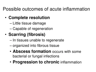
Inflammation may be acute or chronic, depending on the nature of the stimulus and the effectiveness of the initial reaction in eliminating the stimulus or the damaged tissues. Acute inflammation is rapid in onset (typically minutes) and is of short duration, lasting for hours or a few days; its main characteristics are the exudation of fluid and plasma proteins (edema) and the emigration of leukocytes, predominantly neutrophils (also called polymorphonuclear leukocytes). When acute inflammation is successful in eliminating the offenders the reaction subsides, but if the response fails to clear the invaders it can progress to a chronic phase. Chronic inflammation may follow acute inflammation or be insidious in onset. It is of longer duration and is associated with the presence of lymphocytes and macrophages, the proliferation of blood vessels, fibrosis, and tissue destruction.
http://ars.els-cdn.com/content/image/1-s2.0-S000294401060209X-gr1.jpghttp://ars.els-cdn.com/content/image/1-s2.0-S000294401060209X-gr1.jpg http://upload.wikimedia.org/wikipedia/commons/thumb/1/1d/Wintertenen.jpg/250px-Wintertenen.jpg
Inflammation is terminated when the offending agent is eliminated. The reaction resolves rapidly, because the mediators are broken down and dissipated and the leukocytes have short life spans in tissues. Anti-inflammatory mechanisms are activated that serve to control the response and prevent it from causing excessive damage to the host.
Inflammation may be harmful Mechanisms designed to destroy foreign invaders and necrotic tissues have an intrinsic ability to injure normal tissues. When inflammation is inappropriately directed against self tissues or is not adequately controlled, it becomes the cause of injury and disease. In fact, in clinical medicine, great attention is given to the damaging consequences of inflammation. (Inflammatory reactions underlie common chronic diseases, such as rheumatoid arthritis, atherosclerosis, and lung fibrosis, as well as life-threatening hypersensitivity reactions to insect bites, drugs, and toxins. For this reason our pharmacies abound with anti-inflammatory drugs, which ideally would control the harmful sequelae of inflammation yet not interfere with its beneficial effects.)
The inflammatory response is closely intertwined with the process of repair. At the same time as inflammation destroys, dilutes, and walls off the injurious agent, it sets into motion a series of events that try to heal the damaged tissue. Repair begins during inflammation but reaches completion usually after the injurious influence has been neutralized. In the process of repair the injured tissue is replaced through regeneration of native parenchymal cells, by filling of the defect with fibrous tissue (scarring) or, most commonly, by a combination of these two processes. http://arztartem.files.wordpress.com/2011/03/5701178x21.jpg
Acute inflammation has three major components: alterations in vascular caliber that lead to an increase in blood flow. structural changes in the microvasculature that permit plasma proteins and leukocytes to leave the circulation. emigration of the leukocytes from the microcirculation, their accumulation in the focus of injury, and their activation to eliminate the offending agent.
STIMULI FOR ACUTE INFLAMMATION Acute inflammatory reactions may be triggered by a variety of stimuli: • Infections • Tissue necrosis • Foreign bodies • Immune reactions (also called hypersensitivity reactions) All inflammatory reactions share the same basic features.
REACTIONS OF BLOOD VESSELS IN ACUTE INFLAMMATION In inflammation, blood vessels undergo a series of changes that are designed to maximize the movement of plasma proteins and circulating cells out of the circulation and into the site of infection or injury. The escape of fluid, proteins, and blood cells from the vascular system into the interstitial tissue or body cavities is known as exudation. An exudate is an extravascular fluid that has a high protein concentration, contains cellular debris, and has a high specific gravity. http://compepid.tuskegee.edu/syllabi/pathobiology/pathology/genpath/image7.2%20.gif http://1.bp.blogspot.com/-V2p2vHTpgNQ/TkrFpkpGQAI/AAAAAAAAAFM/ZLz0p4O69AI/s1600/Specific_Gravity.jpg
Fig. 1 Specific gravity of blood cells. Rainer Moog Apheresis techniques for collection of peripheral blood progenitor cells Transfusion and Apheresis Science Volume 31, Issue 3 2004 207 - 220 http://dx.doi.org/10.1016/j.transci.2004.09.006
Exudate Its presence implies an increase in the normal permeability of small blood vessels in an area of injury and, therefore, an inflammatory reaction. In contrast, a transudate is a fluid with low protein content (most of which is albumin), little or no cellular material, and low specific gravity. It is essentially an ultrafiltrate of blood plasma that results from osmotic or hydrostatic imbalance across the vessel wall without an increase in vascular permeability. Edema denotes an excess of fluid in the interstitial tissue or serous cavities; it can be either an exudate or a transudate. Pus, a purulent exudate, is an inflammatory exudate rich in leukocytes (mostly neutrophils), the debris of dead cells and, in many cases, microbes. http://www.clinimed.co.uk/Portals/10/images/Figure%2013%20Exudate.jpg http://path.upmc.edu/cases/case146/images/micro7.jpg
Changes in Vascular Flow and Caliber Changes in vascular flow and caliber begin early after injury and consist of the following. Vasodilation is one of the earliest manifestations of acute inflammation; sometimes it follows a transient constriction of arterioles, lasting a few seconds. Vasodilation first involves the arterioles and then leads to opening of new capillary beds in the area. The result is increased blood flow, which is the cause of heat and redness (erythema) at the site of inflammation. Vasodilation is induced by the action of several mediators, notably histamine and nitric oxide (NO), on vascular smooth muscle. Vasodilation is quickly followed by increased permeability of the microvasculature, with the outpouring of protein-rich fluid into the extravascular tissues; this process is described in detail below.
http://www.sciencedirect.com/science/article/pii/S1521689603001162http://www.sciencedirect.com/science/article/pii/S1521689603001162
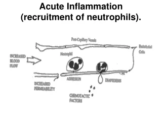
The loss of fluid and increased vessel diameter lead to slower blood flow, concentration of red cells in small vessels, and increased viscosity of the blood. These changes result in dilation of small vessels that are packed with slowly moving red cells, a condition termed stasis, which is seen as vascular congestion (producing localized redness) upon examination of the involved tissue. http://usmlepathslides.tumblr.com/post/18136094023/acute-inflammation-stasis-this-slide-shows-a-small Acute inflammation-stasisThis slide shows a small venule filled with neutrophils and red blood cells. There are also red blood cells outside of the blood vessel which represents diapedesis. Fluid (edema) has also leaked out of the blood vessel which is the cause of “stasis” of the red blood cells and causes the neutrophils to begin to slow their passage through the blood stream. This process of vasodilatation and increased vascular permeability, due to histamine, facilitates the adhesion of the neutrophils to endothelial cells.
As stasis develops, blood leukocytes, principally neutrophils, accumulate along the vascular endothelium. At the same time endothelial cells are activated by mediators produced at sites of infection and tissue damage, and express increased levels of adhesion molecules. Leukocytes then adhere to the endothelium, and soon afterward they migrate through the vascular wall into the interstitial tissue, in a sequence that is described later. http://www.sciencedirect.com/science/article/pii/S1521689603001162
http://www.sciencedirect.com/science/article/pii/S1521689603001162http://www.sciencedirect.com/science/article/pii/S1521689603001162
Increased Vascular Permeability (Vascular Leakage) Contraction of endothelial cells resulting in increased interendothelial spaces is the most common mechanism of vascular leakage and is elicited by histamine, bradykinin, leukotrienes, the neuropeptide substance P, and many other chemical mediators. It is called the immediate transient response because it occurs rapidly after exposure to the mediator and is usually short-lived (15–30 minutes). In some forms of mild injury (e.g. after burns, x-irradiation or ultraviolet radiation, and exposure to certain bacterial toxins), vascular leakage begins after a delay of 2 to 12 hours, and lasts for several hours or even days; this delayed prolonged leakage may be caused by contraction of endothelial cells or mild endothelial damage. Late-appearing sunburn is a good example of this type of leakage.
http://ars.els-cdn.com/content/image/1-s2.0-S0301008209001981-gr1.jpghttp://ars.els-cdn.com/content/image/1-s2.0-S0301008209001981-gr1.jpg
Endothelial injury, resulting in endothelial cell necrosis and detachment. Endothelium damage is encountered in severe injuries, for example, in burns, or by the actions of microbes that target endothelial cells. Neutrophils that adhere to the endothelium during inflammation may also injure the endothelial cells and thus amplify the reaction. In most instances leakage starts immediately after injury and is sustained for several hours until the damaged vessels are thrombosed or repaired.
Increased transport of fluids and proteins, called transcytosis, through the endothelial cell. This process may involve channels consisting of interconnected, uncoated vesicles and vacuoles called the vesiculovacuolar organelle, many of which are located close to intercellular junctions. Certain factors, such as VEGF, seem to promote vascular leakage in part by increasing the number and perhaps the size of these channels. FIGURE 2-3 Principal mechanisms of increased vascular permeability in inflammation, and their features and underlying causes. NO, nitric oxide; VEGF, vascular endothelial growth factor.
http://www.nature.com/nrn/journal/v7/n1/images/nrn1824-f3.jpghttp://www.nature.com/nrn/journal/v7/n1/images/nrn1824-f3.jpg
Responses of Lymphatic Vessels In inflammation, lymph flow is increased and helps drain edema fluid that accumulates due to increased vascular permeability. In addition to fluid, leukocytes and cell debris, as well as microbes, may find their way into lymph. Lymphatic vessels, like blood vessels, proliferate during inflammatory reactions to handle the increased load. The lymphatics may become secondarily inflamed (lymphangitis), as may the draining lymph nodes (lymphadenitis). http://s3.amazonaws.com/itriage/google_image_searches/b29c63ba7ebaa73e6b710c52b57e8e01/original.jpg?1269018580
Inflamed lymph nodes are often enlarged because of hyperplasia of the lymphoid follicles and increased numbers of lymphocytes and macrophages. This constellation of pathologic changes is termed reactive, or inflammatory, lymphadenitis. For clinicians the presence of red streaks near a skin wound is a telltale sign of an infection in the wound. This streaking follows the course of the lymphatic channels and is diagnostic of lymphangitis; it may be accompanied by painful enlargement of the draining lymph nodes, indicating lymphadenitis. http://www.meddean.luc.edu/lumen/meded/pathweb/images/44-10.JPG http://www.healthguidance.org/hgimages/16277Armpitlump.jpg
REACTIONS OF LEUKOCYTES IN INFLAMMATION The most important leukocytes in typical inflammatory reactions are the ones capable of phagocytosis, namely neutrophils and macrophages. These leukocytes ingest and kill bacteria and other microbes, and eliminate necrotic tissue and foreign substances. Leukocytes also produce growth factors that aid in repair. When strongly activated, leukocytes may induce tissue damage and prolong inflammation, because the leukocyte products that destroy microbes and necrotic tissues can also injure normal host tissues.
The processes involving leukocytes in inflammation consist of: their recruitment from the blood into extravascular tissues, recognition of microbes and necrotic tissues, and removal of the offending agent. Recruitment of Leukocytes to Sites of Infection and Injury The journey of leukocytes from the vessel lumen to the interstitial tissue, called extravasation, can be divided into the following steps. 1. In the lumen: margination, rolling, and adhesion to endothelium. Vascular endothelium in its normal, unactivated state does not bind circulating cells or impede their passage. In inflammation the endothelium is activated and can bind leukocytes, as a prelude to their exit from the blood vessels. Migration across the endothelium and vessel wall. Migration in the tissues toward a chemotactic stimulus.
Fig. 1. Cell adhesion molecules at the endothelial-cell borders. (a) In resting endothelial cells homophilic interactions between vascular endothelial-cell specific cadherin [VE-cadherin (shown with five cadherin repeats as green rectangles)] on apposing cells, the immunoglobulin (Ig) gene superfamily molecules platelet–endothelial-cell adhesion molecule-1 [PECAM-1 (with six Ig domains indicated as circles)], junctional adhesion molecule (JAM)-A, -B and -C (with two Ig domains indicated) and CD99 (with O-linked sugars indicated by red ‘whiskers’) are established. (b) Under inflammatory conditions, there might be rearrangement of these molecules within the cell. A combination of the inflammatory cytokines tumor necrosis factor-α (TNF-α) and interferon-γ (IFN-γ) induces redistribution of JAM-A to the endothelial apical surface. The same combination reduces the expression of PECAM-1; IFN-γ alone (but not TNF-α or interleukin-1β) induces partial redistribution of PECAM-1 to the apical surface (not shown). (c) During leukocyte transendothelial migration, the density of VE-cadherin in the membrane adjacent to the advancing leukocyte decreases dramatically while the density of JAM-A and recycling PECAM-1 increases. It is not known whether total PECAM density increases locally or if just the density of the recycling pool increases. http://ars.els-cdn.com/content/image/1-s2.0-S1471490603001170-gr1.gif
Leukocyte Adhesion to Endothelium. In normally flowing blood in venules, red cells are confined to a central axial column, displacing the leukocytes toward the wall of the vessel. Because blood flow slows early in inflammation (stasis), hemodynamic conditions change (wall shear stress decreases), and more white cells assume a peripheral position along the endothelial surface. This process of leukocyte redistribution is called margination. Subsequently, individual and then rows of leukocytes adhere transiently to the endothelium, detach and bind again, thus rolling on the vessel wall. The cells finally come to rest at some point where they adhere firmly (resembling pebbles over which a stream runs without disturbing them). http://img2.tfd.com/mk/I/X2604-I-14.png http://www.pathguy.com/lectures/polys_marginating.jpg
The adhesion of leukocytes to endothelial cells is mediated by complementary adhesion molecules on the two cell types whose expression is enhanced by secreted proteins called cytokines. Cytokines are secreted by cells in tissues in response to microbes and other injurious agents, thus ensuring that leukocytes are recruited to the tissues where these stimuli are present. The initial rolling interactions are mediated by a family of proteins called selectins.
There are three types of selectins: one expressed on leukocytes (L-selectin), one on endothelium (E-selectin), and one in platelets and one on endothelium (P-selectin). The ligands for selectins are sialylated oligosaccharides bound to mucin-like glycoprotein backbones. The expression of selectins and their ligands is regulated by cytokines produced in response to infection and injury. Tissue macrophages, mast cells, and endothelial cells that encounter microbes and dead tissues respond by secreting several cytokines, including tumor necrosis factor (TNF), interleukin-1 (IL-1), and chemokines (chemoattractant cytokines).
http://www.sciencedirect.com/science/article/pii/S1521689603001162http://www.sciencedirect.com/science/article/pii/S1521689603001162
TNF and IL-1 act on the endothelial cells of post-capillary venules adjacent to the infection and induce the coordinate expression of numerous adhesion molecules (Fig. 2-5). Within 1 to 2 hours the endothelial cells begin to express E-selectin and the ligands for L-selectin. Other mediators such as histamine, thrombin, and platelet-activating factor (PAF), stimulate the redistribution of P-selectin from its normal intracellular stores in endothelial cell granules (called Weibel-Palade bodies) to the cell surface. Leukocytes express L-selectin at the tips of their microvilli and also express ligands for E- and P-selectins, all of which bind to the complementary molecules on the endothelial cells. These are low-affinity interactions with a fast off-rate, and they are easily disrupted by the flowing blood. As a result, the bound leukocytes bind, detach, and bind again, and thus begin to roll along the endothelial surface.
FIGURE 2-5 Regulation of expression of endothelial and leukocyte adhesion molecules. A, Redistribution of P-selectin from intracellular stores to the cell surface. B, Increased surface expression of selectins and ligands for integrins upon cytokine activation of endothelium. C, Increased binding avidity of integrins induced by chemokines. Clustering of integrins contributes to their increased binding avidity (not shown). IL-1, interleukin-1; TNF, tumor necrosis factor.
Weak rolling interactions slow down the leukocytes and give them the opportunity to bind more firmly to the endothelium. Firm adhesion is mediated by a family of heterodimeric leukocyte surface proteins called integrins. TNF and IL-1 induce endothelial expression of ligands for integrins, mainly vascular cell adhesion molecule 1 (VCAM-1, the ligand for the VLA-4 integrin) and intercellular adhesion molecule-1 (ICAM-1, the ligand for the LFA-1 and Mac-1 integrins). Leukocytes normally express integrins in a low-affinity state. Meanwhile, chemokines that were produced at the site of injury enter the blood vessel, bind to endothelial cell proteoglycans, and are displayed at high concentrations on the endothelial surface. These chemokines bind to and activate the rolling leukocytes. One of the consequences of activation is the conversion of VLA-4 and LFA-1 integrins on the leukocytes to a high-affinity state.[22] The combination of cytokine-induced expression of integrin ligands on the endothelium and activation of integrins on the leukocytes results in firm integrin-mediated binding of the leukocytes to the endothelium at the site of inflammation. The leukocytes stop rolling, their cytoskeleton is reorganized, and they spread out on the endothelial surface.