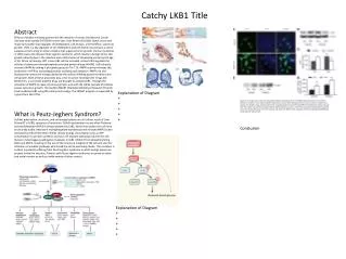
LKB1
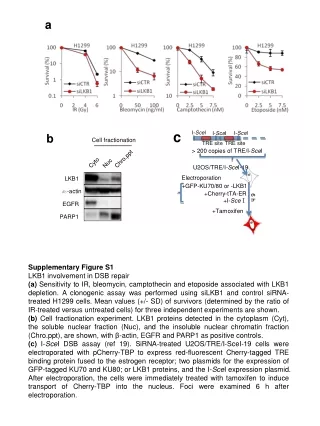
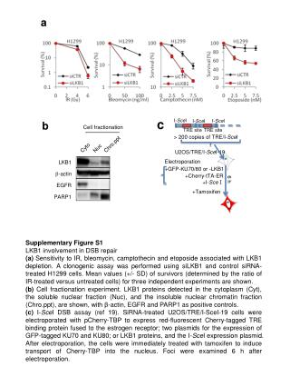
a. I- Sce I. I- Sce I. I- Sce I. TRE site. TRE site. c. b. Cell fractionation. > 200 copies of TRE/I- Sce I. Chro.ppt. Cyto. Nuc. U2OS/TRE/I -Sce I-19. Electroporation. LKB1. +GFP-KU70/80 or -LKB1. b -actin. +Cherry- tTA -ER. 6 h. +I- Sce Ⅰ. EGFR. + Tamoxifen. PARP1.

LKB1
E N D
Presentation Transcript
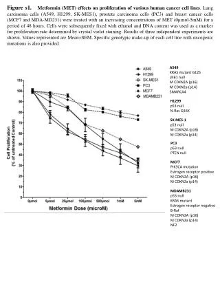
a I-SceI I-SceI I-SceI TRE site TRE site c b Cell fractionation > 200 copies of TRE/I-SceI Chro.ppt Cyto Nuc U2OS/TRE/I-SceI-19 Electroporation LKB1 +GFP-KU70/80 or -LKB1 b-actin +Cherry-tTA-ER 6 h +I-SceⅠ EGFR +Tamoxifen PARP1 Supplementary Figure S1 LKB1 involvement in DSB repair (a) Sensitivity to IR, bleomycin, camptothecin and etoposide associated with LKB1 depletion. A clonogenic assay was performed using siLKB1 and control siRNA-treated H1299 cells. Mean values (+/- SD) of survivors (determined by the ratio of IR-treated versus untreated cells) for three independent experiments are shown. (b) Cell fractionation experiment. LKB1 proteins detected in the cytoplasm (Cyt), the soluble nuclear fraction (Nuc), and the insoluble nuclear chromatin fraction (Chro.ppt), are shown, with β-actin, EGFR and PARP1 as positive controls. (c) I-SceI DSB assay (ref 19). SiRNA-treated U2OS/TRE/I-SceI-19 cells were electroporated with pCherry-TBP to express red-fluorescent Cherry-tagged TRE binding protein fused to the estrogen receptor; two plasmids for the expression of GFP-tagged KU70 and KU80; or LKB1 proteins, and the I-SceI expression plasmid. After electroporation, the cells were immediately treated with tamoxifen to induce transport of Cherry-TBP into the nucleus. Foci were examined 6 h after electroporation.
WCE Flag IP - - - - + + + + Flag-LKB1 Supplementary Figure S2 Interaction between LKB1 and ATM examined by immunoprecipitation (IP) of whole cell extracts (WCE) using anti-FLAG antibody. - - - - + + + + IR Flag-LKB1 BRM ATM
siAMPK2 #2 siCont AMPK2 b-actin Fraction of EGFP positive cells Supplementary Figure S3 Reduced NHEJ activity by AMPK2-depletion using a different set of siRNAs from those used in Figure 3. Results of western blot analysis are also shown.
a siRNA Control siRNA LKB1 siRNA AMPK2 siRNA LKB1+AMPK2 b 2 2 Supplementary Figure S4 Effect of LKB1 and AMPK2 depletion on the number of chromosomal breaks and radials (a) Chromosome breaks and radials in H1299 cells transfected with control, LKB1 and/or AMPK2 siRNA. Slides were Giemsa stained and metaphase spreads were analyzed for the number of chromosomal aberrations. (b) Number of chromosomal aberrations per metaphase spread.
siControl siLKB1 siBRM Supplementary Figure S5 Size fractionation of DNA fragments containing breakpoint junctions. Three major recurrent types of repiatred products (Types I–III in ref 23) were observed all in siControl, siLKB1 and siBRM cells.Experimental details are in ref 23.
WCE Flag IP - - - - + + Flag-LKB1 - - + - - + Flag-LKB1 kd Flag BRM Supplementary Figure S6 WT-LKB1 and kd-LKB1 (SL26) interaction with BRM examined by immunoprecipitaion (IP) of whole cell extract (WCE) using anti-FLAG antibody.
Fraction of EGFP positive cells siAMPK1 siCont AMPK1 Supplementary Figure S7 Reduced NHEJ activity in AMPK1-depleted cells. Results of western blot analysis are also shown. b-actin