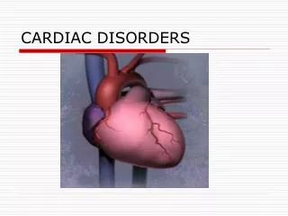
CARDIAC DISORDERS
CARDIAC DISORDERS. Introduction to Cardiac. Every year since 1900 (except 1918) cardiac disease has been the leading cause of death in the US 42 % of all deaths (1 million) are related to CVD Leading cause of death for women 1 in 5 people has experienced and are living with CVD.

CARDIAC DISORDERS
E N D
Presentation Transcript
Introduction to Cardiac • Every year since 1900 (except 1918) cardiac disease has been the leading cause of death in the US • 42 % of all deaths (1 million) are related to CVD • Leading cause of death for women • 1 in 5 people has experienced and are living with CVD
Function of the Heart • Collects deoxygenated blood coming from the body • Sends this blood to the lungs for oxygen • Receives oxygenated blood from lungs • Pumps oxygenated blood and nutrients to the body • Does this an average of 70 times a minute • Pumps 60 mL of blood with each beat or 5 L/min
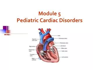
Cardiac Anatomy • 4 chambers • Right atrium- receives blood from the body by way of the superior and inferior vena cava and from the coronary sinuses • Right ventricle- receives blood from the right atrium through the tricuspid valve. It then contracts (systole), and forces blood through the pulmonary valve into the pulmonary artery. The pulmonary artery carries the blood to the lungs and CO2 is released and O2 picked up and transported back to the heart through the pulmonary veins
Cardiac Anatomy, cont. • Left atrium- oxygenated blood received from the lungs and transferred by way of the mitral valve into the left ventricle • Left ventricle- strongest muscular chamber of the heart. Pumps blood through the aortic valve into the aorta
Four Cardiac Valves • Atrioventricular Valves • Open with diastole, close with systole • Tricuspid- between the RA and LA- 3 leaflets • Mitral- between the LA and LV- 2 leaflets • Semilunar valves • Open during systole, close with diastole • Pulmonic- between the RV and the PA • Aortic- between the LV and the aorta
Cardiac Muscle • 3 layers of muscle • Endocardium-inner layer • Lines the heart chambers • Myocardium-middle layer • Muscle fiber which pumps the blood • Epicardium-outer layer • Covering of the heart which contains the coronary arteries
Heart Sounds • S1 • ventricular contraction • Systole • Lub • S2 • ventricular relaxation • Dub • Diastole • Closure of aortic and pulmonic valves
Coronary Arteries • Originate from an area on the aorta beyond the aortic valve • 2 main coronary arteries • RCA- supplies the right atrium, the right ventricle and the inferior of the left ventricle • Left main artery- divides into 2 branches • LAD-supplies most of the front of the LV, and part of the septum • LCX-descends around the back of the LV • Both of these have branches called diagonals and obtuse marginals • Supplied with blood during diastole • Must maintain a mean arterial blood pressure of at least 60 to maintain perfusion of the heart
Conduction System • Composed of special nerve fibers which send impulses throughout the cardiac muscle and initiates a muscle contraction • Cardiac muscle cells possess characteristics of: • Automaticity-can initiate impulses spontaneously • Excitability-cells respond to a stimulus by initiating an impulse • Conductivity-transmit an electrical impulse • Refractoriness – cardiac muscle cannot respond to a stimulus until it has recovered (repolarized) from a pervious stimulus
Conduction System, cont. • Sinoatrial node (SA node) • Major regulator of heart rate • Called the “pacemaker” • Spontaneously initiate impulses at a rate of 60-100, called a sinus rhythm • Atrioventricular node (AV node) • Receives impulse form SA node • Located at the junctional area • Can sent impulse of 40-60 beats without SA node stimulus
Conduction, cont. • Bundle of HIS • Continuation of AV node • Located in the intraventricular septum • Has a right and left branch • Purkinje fibers • Join the Bundle • Terminal branches of system • Can deliver a rate of 20-40 beats a minute • When impulse reaches here ventricles contract
Conduction, cont. • At rest the inside of the cell is negatively charged and outside the cell is positively charged • When an impulse is received from the SA node, there is a change in the electrical charge of the cells – lose their internal negativity and become depolarized – this goes from cell to cell • Repolarization is when the resting state returns • Then the cycle continues
Other Cardiac Stimulus • Autonomic Nervous System • Sympathetic • Fight or flight -^ HR, ^strength of contraction • Parasympathetic (part of the vagus nerve) • Found in the SA and AV nodes • Slows HR • Decreases strength of contraction
Mechanical Function • Phases of Cardiac Cycle • Diastole – 2/3 of cycle, relaxation and filling of the atria and ventricles, “atrial kick” is at the end of this cycle • Systole – contraction and emptying of the ventricles
Mechanical Function, cont. • Stroke volume – is amount of blood ejected from the ventricle with each heart beat. Normal is 60-100 mL • Cardiac output – amount of blood in liters, ejected by the heart each minute. Normal is 4-6 liters/min • To calculate CO = HR x SV
Factors Which Affect Stroke Volume • Preload – determined by the amount of blood returning to the heart from the venous system and the pulmonary system • Afterload – amount of resistance which the heart must overcome to eject blood from the LV into the peripheral vascular system • Contractility – the force of contraction of heart muscle • Inotropic = term which refers to the contractile state of the cell • Positive inotropic • Negative inotropic
O2 Consumption • Determined by the needs of the myocardium • O2 is delivered to the heart muscle by the coronary arteries • 2 ways to increase O2 to the myocardium • Increase coronary blood flow • Increase O2 in the blood by giving O2 supplement