Download
1 / 70
700 likes | 853 Vues
PHYSICAL EXAMINATION OF THE SPINE. Prof. Dr. Şafak Sahir KARAMEHMETOĞLU, MD. İU/CMF/PMRD. CERVICAL SPINE. INSPECTION-1. - Lordosis - Scoliosis - Swelling - Torticollis - Muscle atrophy. INSPECTION-2. - Muscle hypertrophy

E N D
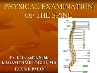
PHYSICAL EXAMINATION OF THE SPINE Prof. Dr. Şafak Sahir KARAMEHMETOĞLU, MD. İU/CMF/PMRD
INSPECTION-1 - Lordosis - Scoliosis - Swelling - Torticollis - Muscle atrophy
INSPECTION-2 - Muscle hypertrophy - Color changes - Arterial pulse - Postural changes
* Bone-Joint - Spinous process - Interspinous space - Foramen PALPATION-1
* SOFT TISSUES - Swelling - PVM - SCMM - Spinalnerves - Ligaments PALPATION-2
* Neighbouring structures - Arterial pulse - Lymph nodules - Thyroid - Trachea - Others PALPATION-3
- Flexion - Extension - Lateral Flexion (45°) - Rotations - Translations - Circumflexion ROM
- Cervical Plexus C1-C4 - Brachial Plexus C5-T1 NEUROLOGIC EXAMINATION
Has no key muscle, flexors, extensors, lateral flexors and rotators are tested in groups. C1: Mainly motor fibres. C2: Key point: protuberantia occipitalis externa. C3: Key point: middle of the fossa supraclavicularis. C4: Key point: acromio-clavicular joint. Cervical Plexus
C5: KM: biceps, brachialis, KP: lateral of the antecubital fossa, DTR: biceps. C6: KM: extensor carpi radialis longus and brevis, KP: middle of the dorsum of the first phalanx of the thumb, DTR: brachioradialis. C7: KM: triceps, KP: middle of the dorsum of the first phalanx of the middle finger, DTR: triceps C8: KM: flexor digitorum profundus, KP: middle of the dorsum of the first phalanx of the little finger. T1:KM: abductor digiti minimi, KP: medial of the antecubital fossa. Brachial Plexus
Distraction: To perform this test, place the open palm of one hand under the patient’s chin, and the other hand under the occiput. Then, gradually lift (distract) to remove its weight from the neck, if the neck and/or arm pain decreases or disappaers, the test is positive. It demonstrates the effect that neck traction might have in relieving pain by widening the foramen, decreasing pressure on the joints capsules around the facet joints. In addition it may help to alleviate muscle spasm by relaxing the contracted muscles. Special Tests -1-
Compression: To perform this test, press down upon the top of the patient’s head while he is either sitting or lying down, if there is an increase in pain in the neck and/or arm(s), then the test is considered to be positive. A narrowing of the neural foramen, pressure on the facet joints or muscle spasm can cause increased pain. In addition, this test may reproduce pain referred to the upper extremity from the cervical helping to locate the neurological level of any existing pathology. Special Tests -2-
Valsalva: To perform this test, have the patient hold his breath and bear down as he/she were moving his/her bowels. Then, ask the patient whether he/she feels any increase in pain, and if so, whether he/she can describes the location. If the response is accurate, the test is positive. This test increases intrathecal pressure. If a space occupying lesion such as a herniated disc or a tumor, is present in the cervical canal, the patient may develop pain in the cervical spine. The pain may also radiate according to the neurological level. Special Tests -3-
Swallowing: Difficultyorpainuponswallowing can sometimes be causedbycervicalspinepathologysuch as bonyprotuberances, osteophytes, orbysofttissueswellingduetohematomas, infection, ortumor in theanteriorportion of thecervicalspine. Special Tests -4-
Adson: To perform this test, take the patient’s radial pulse at the wrist. As you continue to feel the pulse, abduct, extend and externally rotate his arm. Then istruct him/her to take a deep breath and to turn his/her head toward the arm being tested. If there is compression of the subclavian artrey, you will feel a marked diminution or absence of the radial pulse, then the test is positive. This test is used to determine the state of the subclavian artery, which may be compressed by an extra cervical rib or by tightened scalenus anticus and scalenus medius muscles, which can compress the artery where it passes between them on its way to the upper extremity. Special Tests -5-
Spurling: To perform this test, instruct the paient to extend, lateral flex and rotate his/her head. Then, press down upon the the top of the patient’s head while he is either sitting or lying down, if there is an increase in pain in the neck and/or arm(s), then the test is considered to be positive. A narrowing of the neural foramen, pressure on the facet joints or muscle spasm can cause increased pain. In addition, this test may reproduce pain referred to the upper extremity from the cervical helping to locate the neurological level of any existing pathology. Special Tests -6-
Slump: This test is a progressive series of maneuvers designed to place the sciatic nerve roots under increasing tension. The patient sitting on the examining table, flexes the cervical, thoracic and lumbar spine, extends one of the knees and dorsifelexes the foot on the same side. If the patient experiences pain in low-back and/or leg(s), the test is positive. Special Tests -7-
INSPECTION-1 * The patient must be undressed, * Posture * Supine, prone and side-lying
INSPECTION-2 * Spina scapula T3 * End of scapula T7-9 * Medial border of the scapula and spinous processes 5 sm. * End of the ears, acromions and, iliac crests must be horizontal
PALPATION and PRESSION * PVM * Facet joints * Spinous processes * Interspinous spaces
ROM-1 • * Flexion 20-45° • * Extension 20-45° • * Lateral flexion 20-40° • * Rotation 35-50°
ROM-2 * Sitting position, * C7-T12: 3 sm., C7-S1 15 sm. * Structural scoliosis does not change in flexion
Neurologic Examination The level of the lesion by key points There is no key muscle Beevor’s sign
T2: KP: Apex of the axilla T3: KP: Third intercostal space (mid-clavicular line) T4: KP: Fourth intercostal space (nipple level, mid-clavicular line) T5: KP: Fifth intercostal space (mid-clavicular line) T6: KP: Sixth intercostal space (xiphoid level, mid-clavicular line) T7: KP: Between T6 and T8 (mid-clavicular line) T8: KP: Between T7 and T9 (mid-clavicular line) T9: KP: Between T8 and T10 (mid-clavicular line) T10: KP: Umblicus (mid-clavicular line) T11: KP: Between T10 and T12 (mid-clavicular line) T12: KP: Superior of the middle of the inguinal ligament (mid-clavicular line) Key Points
Special tests • * Slump • * Passivescapularapproximation • * Firstthoracalnervestretching
Passive scapular approximation test: The patient lies prone. The shoulders are stretched backwards with the approximation of scapulae. If there is pain or pain worsening the test is pozitive. This test stretches the first thoracal spinal nerve. Special tests - 2
First thoracal nerve stretching test: The patient abducts the shoulder to 90°, flexes the elbow and holds the occipital region of the head and the shoulder is forced to extention. If there is pain or pain worsening the test is pozitive. This test stretches the first thoracal spinal nerve. Special tests - 3
Beevor's sign is the movement of the belly button towards the head on flexing the neck. It is caused by weakness of the lower abdominal muscles. Beevor’s sign is characteristic of spinal cord injury at the T10 level. It has also been described in amyotrophic lateral sclerosis and facioscapulohumeral muscular dystrophy. Special tests - 4
INSPECTION - 1 - Lordosis - Scoliosis - Swelling - Deviation - Muscle atrophy
INSPECTION - 2 - Muscle spasm - Color changes - Lipomas - Abnormal hair - Café au lait spots - Postural changes
PALPATION-1 Bone – Joint: - Spinous processes - Interspinous spaces - Facet joints - İliac crests - Coccyx
PALPATION-2 SOFT TISSUES - Swelling - PVM - Ligaments - Umblicus (L3-4) - Sacral promontorium
PALPATION-3 VALLEIX POINTS: 1. Middle of the trochanter major ve ischial tuberosity 2. Middle of the posteroir thigh 3. Middle of the popliteal fossa 4. Middle of the gastrocnemius muscle 5. Middle of the Achilles tendon