ECG Abnormalities in Electrolyte Imbalances: A Comprehensive Guide
100 likes | 248 Vues
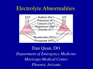
Learn how electrolyte abnormalities affect ECG readings. Understand hyperkalemia, hypokalemia, hypocalcemia, hypercalcemia, and hypomagnesemia, with detailed ECG descriptions and patterns.

ECG Abnormalities in Electrolyte Imbalances: A Comprehensive Guide
E N D
Presentation Transcript
Electrolyte abnormalities and ECG Elias Hanna, MD LSU Cardiology
Hyperkalemia: T wave in hyperkalemia is typically tall and narrow, but does not have to be tall (may be just narrow and peaked pulling ST segment). Tall T means > 2 big boxes in the precordial leads or >1 small box in limb leads, or T wave taller than QRS.
Hypokalemia: ST depression with prominent T (actually U) and prolonged QT when K<2.5-3 Flat T with K~3
Hypokalemia: -T progressively flattens -U wave more and more prominent (looks like T) -ST-segment more and more depressed Large U wave simulates and hides T wave with severe hypokalemia, the ST-T pattern may mimick: ST-segment depression with a flat or upright wide “T” wave (actually U) and a prolonged “QT” interval (actually QTU)
On the other hand, the pattern of T inversion is not seen with hypokalemia:
ECG changes of digoxin effect (digoxin therapy) simulate the changes seen with hypokalemia (U wave and ST depression), except that with digoxin therapy QT is not prolonged
Hypocalcemia: Long QT that is due to a long ST segment, which is different from long QT due to congenital long QT syndrome, drugs, or hypokalemia. T wave is not wide, there is no T wave abnormality.
Hypercalcemia: short QTc <390 ms. No significant ST or T wave abnormality
Hypomagnesemia is not associated with characteristic or specific ECG findings • It is associated with a non-specific prolongation of QT and/or QRS intervals, and is often associated with hypokalemia and hypocalcemia. Therefore, changes related to the latter 2 abnormalities may be seen.