How to perform Routine Anomaly Scan 2008
560 likes | 773 Vues
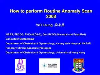
How to perform Routine Anomaly Scan 2008. WC Leung 梁永昌 MBBS, FRCOG, FHKAM(O&G), Cert RCOG (Maternal and Fetal Med) Consultant Obstetrician Department of Obstetrics & Gynaecology, Kwong Wah Hospital, HKSAR Honorary Clinical Associate Professor

How to perform Routine Anomaly Scan 2008
E N D
Presentation Transcript
How to perform Routine Anomaly Scan 2008 WC Leung 梁永昌 MBBS, FRCOG, FHKAM(O&G), Cert RCOG (Maternal and Fetal Med) Consultant Obstetrician Department of Obstetrics & Gynaecology, Kwong Wah Hospital, HKSAR Honorary Clinical Associate Professor Department of Obstetrics & Gynaecology, University of Hong Kong
Ultrasound ScreeningRCOG Working Party 2000 The twenty week anomaly scan is to reassure the woman that her baby appears to have no obvious structural abnormalities. Lack of explicitness about which structures have been examined may lead to confusion. The literature provides a range of detection rates and therefore individual units should provide their own figures to inform women undergoing the "20 week" scan. The value of identifying fetal abnormalities at this stage is that it offers parents options. Some, probably the majority, for serious lesions, will elect to terminate the pregnancy. Those couples who choose to continue the pregnancy have the opportunity to prepare themselves through discussions with health care personnel and self-help groups, whilst attendants can ensure appropriate care during pregnancy and following delivery.
Ultrasound ScreeningRCOG Working Party 2000 Head shape + internal structures, cavum pellucidum, cerebellum, ventricular size at atrium (<10 mm) Face & lips Spine: longitudinal and transverse Abdominal shape and content at level of stomach Abdominal shape and content at level of kidneys and umbilicus Renal pelvis (<5 mm AP measurement) Longitudinal axis - abdominal-thoracic appearance (diaphragm/bladder) Thorax at level of 4 chamber cardiac view Cardiac outflow tracts Arms - three bones and hand (not counting fingers) Legs - three bones and foot (not counting toes)
Head shape + internal structures, cavum pellucidum, cerebellum, ventricular size at atrium (<10 mm) Face & lips Spine: longitudinal and transverse Abdominal shape and content at level of stomach Abdominal shape and content at level of kidneys and umbilicus Renal pelvis (<5 mm AP measurement) Longitudinal axis - abdominal-thoracic appearance (diaphragm/bladder) Thorax at level of 4 chamber cardiac view Cardiac outflow tracts Arms - three bones and hand (not counting fingers) Legs - three bones and foot (not counting toes)
Head shape + internal structures, cavum pellucidum, cerebellum, ventricular size at atrium (<10 mm) Face & lips Spine: longitudinal and transverse Abdominal shape and content at level of stomach Abdominal shape and content at level of kidneys and umbilicus Renal pelvis (<5 mm AP measurement) Longitudinal axis - abdominal-thoracic appearance (diaphragm/bladder) Thorax at level of 4 chamber cardiac view Cardiac outflow tracts Arms - three bones and hand (not counting fingers) Legs - three bones and foot (not counting toes)
Head shape + internal structures, cavum pellucidum, cerebellum, ventricular size at atrium (<10 mm) Face & lips Spine: longitudinal and transverse Abdominal shape and content at level of stomach Abdominal shape and content at level of kidneys and umbilicus Renal pelvis (<5 mm AP measurement) Longitudinal axis - abdominal-thoracic appearance (diaphragm/bladder) Thorax at level of 4 chamber cardiac view Cardiac outflow tracts Arms - three bones and hand (not counting fingers) Legs - three bones and foot (not counting toes)
Head shape + internal structures, cavum pellucidum, cerebellum, ventricular size at atrium (<10 mm) Face & lips Spine: longitudinal and transverse Abdominal shape and content at level of stomach Abdominal shape and content at level of kidneys and umbilicus Renal pelvis (<5 mm AP measurement) Longitudinal axis - abdominal-thoracic appearance (diaphragm/bladder) Thorax at level of 4 chamber cardiac view Cardiac outflow tracts Arms - three bones and hand (not counting fingers) Legs - three bones and foot (not counting toes)
Head shape + internal structures, cavum pellucidum, cerebellum, ventricular size at atrium (<10 mm) Face & lips Spine: longitudinal and transverse Abdominal shape and content at level of stomach Abdominal shape and content at level of kidneys and umbilicus Renal pelvis (<5 mm AP measurement) Longitudinal axis - abdominal-thoracic appearance (diaphragm/bladder) Thorax at level of 4 chamber cardiac view Cardiac outflow tracts Arms - three bones and hand (not counting fingers) Legs - three bones and foot (not counting toes)
Head shape + internal structures, cavum pellucidum, cerebellum, ventricular size at atrium (<10 mm) Face & lips Spine: longitudinal and transverse Abdominal shape and content at level of stomach Abdominal shape and content at level of kidneys and umbilicus Renal pelvis (<5 mm AP measurement) Longitudinal axis - abdominal-thoracic appearance (diaphragm/bladder) Thorax at level of 4 chamber cardiac view Cardiac outflow tracts Arms - three bones and hand (not counting fingers) Legs - three bones and foot (not counting toes)
Head shape + internal structures, cavum pellucidum, cerebellum, ventricular size at atrium (<10 mm) Face & lips Spine: longitudinal and transverse Abdominal shape and content at level of stomach Abdominal shape and content at level of kidneys and umbilicus Renal pelvis (<5 mm AP measurement) Longitudinal axis - abdominal-thoracic appearance (diaphragm/bladder) Thorax at level of 4 chamber cardiac view Cardiac outflow tracts Arms - three bones and hand (not counting fingers) Legs - three bones and foot (not counting toes)
Head shape + internal structures, cavum pellucidum, cerebellum, ventricular size at atrium (<10 mm) Face & lips Spine: longitudinal and transverse Abdominal shape and content at level of stomach Abdominal shape and content at level of kidneys and umbilicus Renal pelvis (<5 mm AP measurement) Longitudinal axis - abdominal-thoracic appearance (diaphragm/bladder) Thorax at level of 4 chamber cardiac view Cardiac outflow tracts Arms - three bones and hand (not counting fingers) Legs - three bones and foot (not counting toes)
Head shape + internal structures, cavum pellucidum, cerebellum, ventricular size at atrium (<10 mm) Face & lips Spine: longitudinal and transverse Abdominal shape and content at level of stomach Abdominal shape and content at level of kidneys and umbilicus Renal pelvis (<5 mm AP measurement) Longitudinal axis - abdominal-thoracic appearance (diaphragm/bladder) Thorax at level of 4 chamber cardiac view Cardiac outflow tracts Arms - three bones and hand (not counting fingers) Legs - three bones and foot (not counting toes)
Sonographic "markers" for aneuploidy Choroid plexus cyst Ventriculomegaly (> 10 mm at the atrium) Echogenic bowel (equivalent to bone density) Head shape Nuchal pad (> 5 mm at 20 weeks) Cysterna magna Echogenic foci in heart Dilated renal pelvis (> 5 mm AP)Short femur/humerus Talipes Sandal gap Clinodactyly Clenched hand Two vessel cord
Evaluation of Routine Obstetric Ultrasound Examination in detecting Fetal Structural Abnormalities in Low Risk Pregnancies WC Leung, CP Lee, MHY Tang Department of Obstetrics & Gynaecology, Tsan Yuk Hospital, The University of Hong Kong, Hong Kong, China Objective:To evaluate routine obstetric ultrasound examination in detecting fetal structural abnormalities in low risk pregnancies. Method:A prospective study of the results of routine obstetric ultrasound examination during the first 19 months after the introduction of this service in a local teaching hospital was performed. Results:3288 women had routine ultrasound examination. Pregnancy outcome could be traced in 3187 women. Fetal ultrasound abnormalities were suspected in 73 cases (2.3% ). Follow-up scans showed the same abnormalities in 26 cases (0.8%). These were confirmed after delivery or abortion in 21 cases (0.7%). The most common fetal abnormality detected by routine ultrasound was dilated renal pelvis (10/21= 47.6%). Major abnormalities detected included: hydrocephalus (2), encephalocoele (1), holoprosencephaly with complex congenital heart disease (1), cystic hygroma (1), truncus arteriosus (1), hypoplastic right heart (1) and diaphragmatic hernia (1). Fetal structural abnormalities were missed by routine ultrasound in 45 cases (1.4%). Majority were cardiac abnormalities (19/45 = 42.2%). Conclusion:The sensitivity of routine ultrasound in detecting fetal structural abnormalities was 31.8%. The specificity was 99.8%. 47 women (1.5%) were potentially subjected to unnecessary anxiety because of suspected fetal abnormalities which were not confirmed or were assessed as insignificant on subsequent scan. (HKJGOM 2000; 1:28-32)
Evaluation of Routine Obstetric Ultrasound Examination in detecting Fetal Structural Abnormalities in Low Risk Pregnancies WC Leung, CP Lee, MHY Tang Department of Obstetrics & Gynaecology, Tsan Yuk Hospital, The University of Hong Kong, Hong Kong, China Objective:To evaluate routine obstetric ultrasound examination in detecting fetal structural abnormalities in low risk pregnancies. Method:A prospective study of the results of routine obstetric ultrasound examination during the first 19 months after the introduction of this service in a local teaching hospital was performed. Results:3288 women had routine ultrasound examination. Pregnancy outcome could be traced in 3187 women. Fetal ultrasound abnormalities were suspected in 73 cases (2.3%). Follow-up scans showed the same abnormalities in 26 cases (0.8%). These were confirmed after delivery or abortion in 21 cases (0.7%). The most common fetal abnormality detected by routine ultrasound was dilated renal pelvis (10/21= 47.6%). Major abnormalities detected included: hydrocephalus (2), encephalocoele (1), holoprosencephaly with complex congenital heart disease (1), cystic hygroma (1), truncus arteriosus (1), hypoplastic right heart (1) and diaphragmatic hernia (1). Fetal structural abnormalities were missed by routine ultrasound in 45 cases (1.4%). Majority were cardiac abnormalities (19/45 = 42.2%). Conclusion:The sensitivity of routine ultrasound in detecting fetal structural abnormalities was 31.8%. The specificity was 99.8%. 47 women (1.5%) were potentially subjected to unnecessary anxiety because of suspected fetal abnormalities which were not confirmed or were assessed as insignificant on subsequent scan. (HKJGOM 2000; 1:28-32) Outdated