Download
1 / 12
120 likes | 285 Vues
Liquifaction Method and Extent of Posterior Capsule Opacification : Two-Year Follow -up. Marie Kalfertova , Mariya Burova, Pavel Rozsival, Nada Jiraskova Department of Ophthalmology, Charles University in Prague, University Hospital in Hradec Kralove, Czech Republic

E N D
LiquifactionMethod and ExtentofPosteriorCapsuleOpacification: Two-YearFollow-up Marie Kalfertova, Mariya Burova, Pavel Rozsival, Nada Jiraskova Department of Ophthalmology, Charles University in Prague, University Hospital in Hradec Kralove, Czech Republic The authors have no financial interest in the subject of this poster Supported in part by Charles University Grant Agency, No. 103809
Purpose • posterior capsule opacification (PCO) is still one of the most common complication following cataract surgery with IOL implantation • we evaluate the extent of PCO after cataract surgery • Right eye: torsional phacoemulsification and AquaLase removal of the epithelial cells • Left eye: torsional phacoemulsification without AquaLase • we examine the safety of this method for intraocular tissue – follow endothelial cell count (ECC), pachymetry
Methods • prospective clinical study • we examine patients 3, 6, 12 and 24 months after surgery, digital retroillumination photographs of the anterior segment, pachymetry, endothelial cell count (ECC) and best corrected visual acuity are obtained • ECC and pachymetry were obtained preoperatively too Evaluation of posterior capsule opacification - • EPCO 2000 software (Evaluation of Posterior Capsule Opacification) • OSCA software (Open-Access Systematic Capsule Assessment) • the comparison of PCO between right eye (with liquifaction method) and left eye is done: paired t test analysis
Methods Liquifaction Method: • uses warm pulses (57°C) of balanced salt solution (BSS) to strain and dissolve the lens for aspiration • we use the pulses for removal of the epithelial cells • within the AquaLase handpiece, 4µL fluid pulses are generated as current passes between electrodes • the BSS pulses are delivered at a maximum rate of 50 Hz
Methods - patients • 50 patients (meanage 69.66 ± 9.1 years) • 34 women (70.68 ± 7.5 years) • 16 men (67.5 ± 11.5 years) • patientswithbilateralcataract, preferablywithsimilardensitygrades • patientswithcorneal and retinaldiseasewereexcluded • informedconsentwasobtainedfromallselectedpatients • twoyearsaftersurgerywere 48 patientsexamined (twopatientshaven´tunderwentthe last examination)
EPCO 2000 - Evaluation of Posterior Capsule Opacification software computer-assisted system of PCO morphologic assessment, introduced in 1997 by Tetz incorporates planimetric and grading assessment the density of the opacification is graded clinically from 0 to 4 the selection process and grading of areas are subjective OSCA system - Open-Access Systematic Capsule Assessment is based on location-sensitive entropy-based texture analysis presented by Aslam (2006) most objective system possible OSCA scores range from 0 (no PCO) to approximately 15 (expected maximum PCO) typical OSCA value for images with very little or no PCO is approximately 0.5 Methods Nekolova J, Pozlerova J, Jiraskova N et al.: Comparison of posterior capsule opacification after two different surgical methods of cataract extraction. Am J Ophthalmol 2008;145:493-498
Methods digital retroillumination photographs after EPCO 2000 evaluation
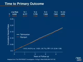
Results Right eye: 0.260 ± 0.198 (3M); 0.259 ± 0.173 (6M); 0.308 ± 0.19 (12M); 0.419 ± 0.252 (24M) Left eye: 0.279 ± 0.170 (3M); 0.280 ± 0.153 (6M); 0.333 ± 0.197 (12M); 0.480 ± 0.313 (24M)
Results Right eye: 0.599 ± 0.240 (3M); 0.605 ± 0.333 (6M); 0.598 ± 0.256(12M); 0.655 ± 0.402 (24M) Left eye: 0.627 ± 0.403 (3M); 0.635 ± 0.360 (6M), 0.629 ± 0.328 (12M), 0.654 ± 0.452 (24M)
Results • there was minimal loss of endothelial cell count and minimal changes in the corneal thickness in both eyes • there was no statistical significant difference between right eye and left eye
Results • Nd-YAG laser capsulotomy one year after surgery underwent one patient (both eyes) and one patient two years postoperatively (right eye) • BCVA two years postoperatively: Right eye: 0.896 ± 0.13 (min.0.6; max.1.2) Left eye: 0.883 ± 0.16 (min.0.4; max.1.2) • ECC (cells/mm²): Right eye - preop.: 2537.6; 3 months postop.: 2354.6 Left eye - preop.: 2582.5; 3 months postop.: 2387.0 • Pachymetry (µm): Right eye – preop.: 565.1; 3 months postop. 561.2 Left eye – preop.: 562.2; 3 months postop. 557.1 • P-values: • EPCO: 0.1 (3M); 0.29 (6M); 0.052 (12M); 0.153 (24M) • OSCA: 0.379 (3M); 0.525 (6M); 0.952 (12M); 0.949 (24M)
Conclusions • an increase in EPCO 2000 indexes and OSCA scores found during 24 months in both eyes • there was no proven significant difference between right eye and left eye, the mean total EPCO index and OSCA score was slightly better for the right eye • our results show that this method is safe for intraocular tissue, our work confirmed previous results Jiraskova et al. Jiraskova N, Kadlecova J, Rozsival P et al. Comparison of the effect of AquaLase and NeoSoniX phacoemulsification on the corneal endothelium. J Cataract Refract Surg 2008; 34:377-382